
Page 1341 - Hematology_ Basic Principles and Practice ( PDFDrive )
P. 1341
C H A P T E R 73
THE PATHOLOGIC BASIS FOR THE CLASSIFICATION OF
NON-HODGKIN AND HODGKIN LYMPHOMAS
Elaine S. Jaffe, Stefania Pittaluga, and John Anastasi
lymph nodes as an incidental finding, and appear to represent a tissue
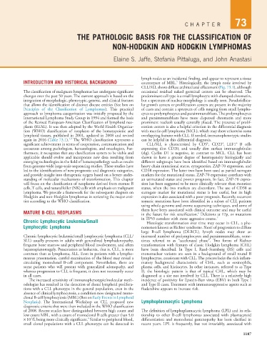
INTRODUCTION AND HISTORICAL BACKGROUND counterpart of MBL. Histologically, the lymph node involved by
5
CLL/SLL shows diffuse architectural effacement (Fig. 73.1), although
The classification of malignant lymphomas has undergone significant occasional residual naked germinal centers can be observed. The
changes over the past 50 years. The current approach is based on the predominant cell type is a small lymphocyte with clumped chromatin,
integration of morphologic, phenotypic, genetic, and clinical features but a spectrum of nuclear morphology is usually seen. Pseudofollicu-
that allows the identification of distinct disease entities (See box on lar growth centers or proliferation centers are present in the majority
Principles of the Classification of Lymphomas). This practical of cases and contain a spectrum of cells ranging from small lympho-
approach to lymphoma categorization was initially proposed by the cytes to prolymphocytes and paraimmunoblasts. The prolymphocytes
International Lymphoma Study Group in 1994 and formed the basis and paraimmunoblasts have more dispersed chromatin and more
of the Revised European-American Classification of lymphoid neo- prominent nucleoli usually centrally placed. The presence of prolif-
plasm (REAL). It was then adopted by the World Health Organiza- eration centers is also a helpful criterion in the differential diagnosis
tion (WHO) classification of neoplasm of the hematopoietic and with mantle cell lymphoma (MCL), which may show otherwise some
lymphoid tissues, published in 2001, updated in 2008 and revised overlapping features with CLL. If needed, immunophenotypic studies
1,2
again in 2016 (Table 73.1). The WHO classification represents a can be helpful in this differential diagnosis.
+
+
+
significant achievement in terms of cooperation, communication and CLL/SLL is characterized by CD5 , CD23 , LEF1 B cells
consensus among pathologists, hematologists, and oncologists. Fur- expressing dim CD20, and usually dim surface immunoglobulin
thermore, it recognizes that any classification system to be viable and (sIg). Cyclin D1 is negative, in contrast to MCL. CLL has been
applicable should evolve and incorporate new data resulting from shown to have a greater degree of heterogeneity biologically and
emerging technologies in the field of hematopathology such as results different subgroups have been identified based on immunoglobulin
from genome-wide large-scale sequencing studies. These studies have heavy chain mutational status, cytogenetics, ZAP-70 expression, and
led to the identifications of new prognostic and diagnostic categories, CD38 expression. The latter two have been used as partial surrogate
and provide insight into therapeutic targets based on a better under- markers for the mutational status. ZAP-70 expression correlates with
standing of molecular mechanisms of transformation. This chapter an unmutated status and poorer prognosis. In fact, ZAP-70 expres-
will focus on the classification of neoplasms derived from mature B sion has been suggested to be more clinically relevant than mutation
cells, T cells, and natural killer (NK) cells with emphasis on malignant status, when the two markers are discordant. The use of CD38 as
lymphoma. We provide a framework for the subsequent chapters on surrogate marker for mutational status is less useful, but its high
Hodgkin and non-Hodgkin lymphomas in reviewing the major enti- expression is also associated with a poor prognosis. Recently, recurrent
ties according to the WHO classification. somatic mutations have been identified in a subset of CLL patients
using whole-genome and exome sequencing techniques, and some of
MATURE B-CELL NEOPLASMS them have been associated with clinical outcome and may be useful
6
in the future for risk stratification. Deletions at 17p, or mutations
Chronic Lymphocytic Leukemia/Small in TP53 correlate with more aggressive course. 7
Histologic transformation over time may occur in CLL, a phe-
Lymphocytic Lymphoma nomenon known as Richter syndrome. Short of progression to diffuse
large B-cell lymphoma (DLBCL), lymph nodes may show an
Chronic lymphocytic leukemia/small lymphocytic lymphoma (CLL/ increased number of prolymphocytes and paraimmunoblasts, some-
SLL) usually presents in adults with generalized lymphadenopathy, times referred to as “accelerated phase”. Two forms of Richter
frequent bone marrow and peripheral blood involvement, and often transformation with features of classic Hodgkin lymphoma (CHL)
hepatosplenomegaly. Presentation as leukemia, that is, CLL, is more have been described. In Type I, Reed–Sternberg (RS) cells and
common than as lymphoma, SLL. Even in patients with a lympho- mononuclear variants are seen in a background of small round B
matous presentation, careful examination of the blood may reveal a lymphocytes, consistent with CLL. The process lacks the rich inflam-
circulating monoclonal B-cell component. Nevertheless, there are matory background characteristic of CHL, such as eosinophils,
some patients who will present with generalized adenopathy, and plasma cells, and histiocytes. In other instances, referred to as Type
whereas progression to CLL is frequent, it does not necessarily occur II, the histologic pattern is that of typical CHL, which may be
in all cases. diagnosed at a site not involved by CLL. There is a relatively high
The increased sensitivity of immunophenotypic/molecular meth- incidence of positivity for Epstein-Barr virus (EBV) in both Type I
odologies has resulted in the detection of clonal lymphoid prolifera- and Type II cases. Treatment with immunosuppressive agents such as
tions with a CLL phenotype in the general population, even in the fludarabine appears to increase risk.
absence of clinical lymphocytosis, a condition now designated mono-
clonal B-cell lymphocytosis (MBL) (Box on Early Events in Lymphoid
Neoplasia). The International Workshop on CLL proposed new Lymphoplasmacytic Lymphoma
diagnostic criteria that were then included in the WHO classification
of 2008. Recent studies have distinguished between high count and The definition of lymphoplasmacytic lymphoma (LPL) and its rela-
low count MBL, with a count of monoclonal B cells greater than 5.0 tionship to other B-cell lymphomas associated with plasmacytoid
3
4
× 10 /L being more clinically significant. Similar to peripheral blood, differentiation and monoclonal gammopathy has been clarified in
small clonal populations with a CLL phenotype can be detected in recent years. LPL is frequently, but not invariably, associated with
1187