
Page 1342 - Hematology_ Basic Principles and Practice ( PDFDrive )
P. 1342
1188 Part VII Hematologic Malignancies
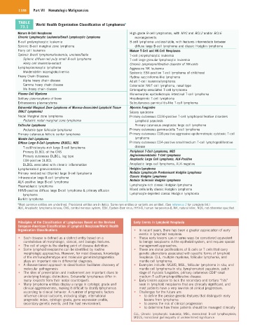
TABLE World Health Organization Classification of Lymphomas a
73.1
Mature B-Cell Neoplasms High grade B-cell lymphomas, with MYC and BCL2 and/or BCL6
Chronic Lymphocytic Leukemia/Small Lymphocytic Lymphoma rearrangements
B-cell prolymphocytic leukemia B-cell lymphoma unclassifiable, with features intermediate between
Splenic B-cell marginal zone lymphoma diffuse large B-cell lymphoma and classic Hodgkin lymphoma
Hairy cell leukemia Mature T-Cell and NK-Cell Neoplasms
Splenic B-cell lymphoma/leukemia, unclassifiable T-cell prolymphocytic leukemia
Splenic diffuse red pulp small B-cell lymphoma T-cell large granular lymphocytic leukemia
Hairy cell leukemia-variant Chronic lymphoproliferative disorder of NK-cells
Lymphoplasmacytic lymphoma Aggressive NK leukemia
Waldenström macroglobulinemia Systemic EBV-positive T-cell lymphoma of childhood
Heavy Chain Diseases Hydroa vacciniforme-like lymphoma
Alpha heavy chain disease Adult T-cell leukemia/lymphoma
Gamma heavy chain disease Extranodal NK/T-cell lymphoma, nasal type
Mu heavy chain disease Enteropathy-associated T-cell lymphoma
Plasma Cell Myeloma Monomorphic epitheliotropic intestinal T-cell lymphoma
Solitary plasmacytoma of bone Hepatosplenic T-cell lymphoma
Extraosseous plasmacytoma Subcutaneous panniculitis-like T-cell lymphoma
Extranodal Marginal Zone Lymphoma of Mucosa-Associated Lymphoid Tissue Mycosis Fungoides
(MALT Lymphoma) Sézary syndrome
Nodal Marginal zone lymphoma Primary cutaneous CD30-positive T-cell lymphoproliferative disorders
Pediatric nodal marginal zone lymphoma Lymphoid papulosis
Follicular Lymphoma Primary cutaneous anaplastic large cell lymphoma
Pediatric-type follicular lymphoma Primary cutaneous gamma-delta T-cell lymphoma
Primary cutaneous follicle center lymphoma Primary cutaneous CD8-positive aggressive epidermotropic cytotoxic T-cell
Mantle Cell Lymphoma lymphoma
Diffuse Large B-Cell Lymphoma (DLBCL), NOS Primary cutaneous CD4-positive small/medium T-cell lymphoproliferative
T-cell/histiocyte–rich large B-cell lymphoma disease
Primary DLBCL of the CNS Peripheral T-Cell Lymphoma, NOS
Primary cutaneous DLBCL, leg type Angioimmunoblastic T-Cell Lymphoma
EBV-positive DLBCL Anaplastic Large Cell Lymphoma, ALK-Positive
DLBCL associated with chronic inflammation Anaplastic large cell lymphoma, ALK-negative
Lymphomatoid granulomatosis Hodgkin Lymphoma
Primary mediastinal (thymic) large B-cell lymphoma Nodular Lymphocyte Predominant Hodgkin Lymphoma
Intravascular large B-cell lymphoma Classic Hodgkin Lymphoma
ALK-positive large B-cell lymphoma Nodular Sclerosis Hodgkin Lymphoma
Plasmablastic lymphoma Lymphocyte-rich classic Hodgkin lymphoma
HHV8-positive diffuse large B-cell lymphoma & primary effusion Mixed cellularity classic Hodgkin lymphoma
lymphoma Lymphocyte-depleted classic Hodgkin lymphoma
Burkitt lymphoma
a Most common entities are underlined. Provisional entities are in italics. Some rare entities or variants are omitted. (See reference 2 for complete list.)
ALK, Anaplastic lymphoma kinase; CNS, central nervous system; EBV, Epstein-Barr virus; HHV-8, human herpesvirus-8; NK, natural killer; NOS, not otherwise specified.
Principles of the Classification of Lymphomas Based on the Revised Early Events in Lymphoid Neoplasia
European-American Classification of Lymphoid Neoplasm/World Health
Organization Classifications • In recent years, there has been a greater appreciation of early
events in lymphoid neoplasia.
• Each disease is defined as a distinct entity based on a • These early lesions can in some ways be considered equivalent
constellation of morphologic, clinical, and biologic features. to benign neoplasms in the epithelial system, and require special
• The cell of origin is the starting point of disease definition. management approaches.
• Some lymphoid neoplasms can be identified by routine • These are clonal proliferations of B cells or T cells that carry
morphologic approaches. However, for most diseases, knowledge genetic aberrations associated with specific forms of lymphoid
of the immunophenotype and molecular genetics/cytogenetics neoplasia: CLL, multiple myeloma, follicular lymphoma, and
plays an important role in differential diagnosis. mantle cell lymphoma.
• A disease-based approach to classification facilitates discovery of • Examples include: MGUS, MBL, follicular lymphoma in situ, and
molecular pathogenesis mantle cell lymphoma in situ; lymphomatoid papulosis, patch
+
• The sites of presentation and involvement are important clues to stage of mycosis fungoides, primary cutaneous CD4 small
underlying biologic distinctions. Extranodal lymphomas differ in medium T-cell lymphoproliferative disease.
many respects from their nodal counterparts. • Early lesions appear to lack the secondary and tertiary “hits”
• Many lymphoma entities display a range in cytologic grade and seen in lymphoid neoplasms that are clinically significant, and
clinical aggressiveness, making it difficult to stratify lymphomas most patients have a very low risk of clinical progression.
according to clinical behavior. A number of prognostic factors • Challenges for the future are:
influence clinical outcome, including stage, international • to define the precise genetic features that distinguish early
prognostic index, cytologic grade, gene expression profile, lesions from lymphoma
secondary genetic events, and the host environment. • to assess the risk of clinical progression
• to determine how these patients should be managed clinically
CLL, Chronic lymphocytic leukemia; MBL, monoclonal B-cell lymphocytosis;
MGUS, monoclonal gammopathy of undetermined significance.