
Page 1346 - Hematology_ Basic Principles and Practice ( PDFDrive )
P. 1346
1192 Part VII Hematologic Malignancies
A A B B CC
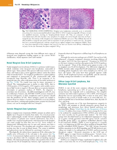
Fig. 73.5 MARGINAL ZONE LYMPHOMA. Marginal zone lymphomas commonly occur at extranodal
sites arising from mucosa-associated lymphoid tissue (MALT). MALT lymphomas typically infiltrate or invade
into epithelial structures, resulting in “lymphoepithelial lesions” (A). They are composed of small to
intermediate-sized cells with abundant clear cytoplasms (A, detail). The normal lymph node does not have a
marginal zone, but primary nodal marginal zone lymphomas (NMZL) can occur. They infiltrate the node in
what would be a marginal zone pattern with an expansion of cells peripheral to mantle zone (B). The spleen
does have a normal marginal zone, and this can give rise to a splenic marginal zone lymphoma (SMZL). Early
on, these show expansion of the marginal zone areas (C) but later can become more diffuse, infiltrating the
red pulp. In the case illustrated, the spleen weighed 1700 g.
differences were detected among the three different main types of frequently observed. Progression to diffuse large B-cell lymphoma can
marginal zone lymphomas lending support to the current WHO be seen.
classification, which separates these three entities. 14 Although the molecular pathogenesis of SMZL has not been fully
delineated, a frequent cytogenetic alteration involving deletions of
15
the region 7q(22-32) has been reported. Mutations in NOTCH2
Nodal Marginal-Zone B-Cell Lymphoma are the most common event but other genes in the NOTCH pathway
16
may be targeted. Many of the affected genes appear to play a role
Nodal marginal zone lymphoma (NMZL) is a primary nodal disease, in marginal zone B-cell development. The differential diagnosis of
which resembles other marginal zone lymphomas, extranodal or SMZL includes other unspecified B-cell lymphomas of the spleen,
splenic types. These patients often present with bone marrow involve- including splenic lymphoma with villous lymphocytes (SLVL), and
ment, and tend to have a more aggressive clinical course than those hairy cell variant. The latter have been grouped together under
with extranodal MALT. The neoplastic proliferation is polymorphous splenic B-cell lymphoma/leukemia unclassifiable, and the interrela-
and composed of monocytoid B cells, plasmacytoid cells, with tionship among these disorders is not fully resolved.
interspersed large blast-like cells. There is an expansion of the
marginal-zone area, often with preservation of the nodal architecture
(see Fig. 73.5B). The mantle zone may be intact, attenuated, or Diffuse Large B-Cell Lymphoma, Not
effaced. The immunophenotype is similar to other MZL, that is, Otherwise Specified
CD20-positive, CD10-negative, CD5-negative, with variable expres-
sion of IgD (weak to negative). Because there are no precise immuno- DLBCL is one of the more common subtypes of non-Hodgkin
phenotypic or genotypic markers of NMZL, the diagnosis is lymphoma, representing up to 40% of cases. It has an aggressive
sometimes one of exclusion. The differential diagnosis with LPL may natural history but responds well to chemotherapy. The complete
be problematic; however, the MYD88 (L265P) somatic mutation is remission rate with modern regimens is 75% to 80%, with long-term
detected infrequently in MZL lymphomas and its presence should disease-free survival approaching 50% or more in most series. This
raise the possibility of LPL. More stringent criteria are needed to lymphoma may present in lymph nodes or in extranodal sites. Fre-
separate these two entities. A variant of nodal MZL occurs in children; quent extranodal sites of involvement include bone, skin, thyroid, GI
these cases show a striking male predominance, present with localized tract, and lung.
disease, and can be managed with local therapies. 1 DLBCL represents one of the most heterogeneous categories in
the WHO, and attempts to identify prognostic groups based on
morphology and phenotype have shown limited usefulness and
Splenic Marginal-Zone Lymphoma reproducibility (Boxon Varied Basis for the Recognition of Diverse
Entities). To address these issues, DLBCLs were among the first cases
Splenic marginal-zone lymphoma (SMZL) presents in adults and is to be analyzed by complementary DNA (cDNA) array technology,
17
slightly more frequent in females than males. The clinical presenta- and more recently also by genome-wide analysis. By GEP three
tion is splenomegaly, usually without peripheral lymphadenopathy. groups were identified based on the differential expression of a large
The majority of patients have marrow involvement, but there is set of genes, namely germinal center-like group (GCB), activated
usually only a modest lymphocytosis, with elevations in the lympho- B-cell–like group (ABC), and primary mediastinal (thymic) large
cyte count usually less than that seen in CLL. Some evidence of B-cell lymphoma (PMBL). PMBL is now recognized as a separate
plasmacytoid differentiation may be seen and patients may have a entity, and adaptations in GEP now allow profiling of formalin fixed
18
small M component. The abundant pale cytoplasm evident in tissue paraffin-embedded (FFPE) biopsies. The ABC subtype frequently
sections may also be seen in blood smears. The course is indolent, exhibits mutations in the BCR-signaling and NF-κB pathways pro-
and splenectomy may be followed by a prolonged remission. viding new insight in the pathogenesis of DLBCL and new potential
19
Histologically, the spleen shows expansion of the white pulp, but therapeutic targets. Recurrent mutations in the GCB type of
17
usually some infiltration of the red pulp is also present (see Fig. DLBCL appear to target histone-modifying genes. Somatic muta-
73.5C). A characteristic biphasic pattern in the neoplastic white pulp tions in EZH2 also have been identified in FL, another tumor of
has been described, with the neoplastic cells surrounding regressed germinal center derivation.
follicles. The immunophenotype of these cells resembles that of other DLBCLs are composed of large, transformed lymphoid cells with
marginal-zone B-cell lymphomas; however, IgD expression is more nuclei at least twice the size of a small lymphocyte (Fig. 73.6). The