
Page 1343 - Hematology_ Basic Principles and Practice ( PDFDrive )
P. 1343
Chapter 73 The Pathologic Basis for the Classification of Non-Hodgkin and Hodgkin Lymphomas 1189
A BB CC D D E
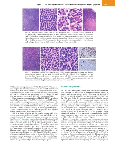
Fig. 73.1 SMALL LYMPHOCYTIC LYMPHOMA. Low-power view (A) illustrates a diffuse effacement of
the lymph node. A monotonous population of small lymphocytes is seen at higher power (B). These have
fairly round nuclear contours, condensed nuclear chromatin, and inconspicuous or absent nucleoli. Only rare
larger cells are present. Small lymphocytic lymphoma can transform to large cell lymphoma (C) and occasion-
ally to Hodgkin lymphoma (D). Patients can also develop worsening lymphadenopathy from viral infections
such as herpes simplex virus, in which the node typically shows focal necrosis (E).
Kappa
Lambda
A A BB C C D D
Fig. 73.2 LYMPHOPLASMACYTIC LYMPHOMA (LPL). Lymphoplasmacytic lymphoma and Walden-
ström macroglobulinemia have nearly identical morphology. There is a diffuse infiltrate (A) of small lympho-
cytes that have plasmacytoid features or interspersed plasma cells ((B), bone marrow; (C), lymph node).
Intranuclear inclusions can sometimes be seen. Evaluation for κ and λ by immunohistochemical stains can
demonstrate clonality in the plasma cells and plasmacytoid lymphocytes (D).
Waldenström macroglobulinemia (WM). The 2008 WHO classifica- Mantle Cell Lymphoma
tion adopted the approach advocated at the second international
workshop on WM, which defined WM as the presence of an, immu- MCL is a distinct entity that has been more precisely defined in recent
noglobulin M (IgM) monoclonal gammopathy of any concentration years through the integration of immunophenotypic, molecular
3
8
associated with bone marrow involvement by LPL. Hence LPL and genetic, and clinicopathologic studies. The molecular hallmark of
WM are not synonymous, with WM defying a subset of LPL. MCL is the t(11;14)(q13;q32) involving Cyclin D1 (CCND1) and
More recently recurrent mutations in MYD88 (L265P) have been the IGH gene. Cyclin D1 overexpression is believed to be essential
identified in greater than 90% of WM patients and are highly associ- in the pathogenesis of MCL. However, rare variants negative for
ated with LPL, but infrequently seen in other B-cell lymphomas with Cyclin D1 with similar immunomorphology and gene expression
9
plasmacytoid features, such as marginal zone lymphomas. In addi- signature have been identified, and most often have translocations
tion, nonsense and frameshift mutations involving CXC-chemokine involving CCND2. Sox11 is overexpressed in most Cyclin D1 posi-
10
receptor 4, similar to those occurring in warts hypogammaglobu- tive and negative cases. The postulated normal counterpart is the
+
+
+
linemia infections myelokathexis (WHIM) patients, were reported CD5 “naive” B cell, sIgM and sIgD , which can be found in the
and are associated with heavy disease burden. peripheral blood and in the mantle of reactive germinal centers.
LPL is a disease of adult life that usually presents with bone Mutational analysis of the rearranged immunoglobulin variable
marrow involvement, and sometimes with nodal and splenic involve- region genes shows few or no somatic mutations; however, similarly
ment (splenomegaly), vague constitutional symptoms, and anemia. to CLL a subset of MCL have mutated IG genes.
(see Chapter 87) The tumor consists of a diffuse proliferation of small Recently, because of the widespread use of immunohistochemistry,
lymphocytes, plasmacytoid lymphocytes, and plasma cells, with or early involvement of lymph node by cells carrying t(11;14) transloca-
without Dutcher bodies (Fig. 73.2). The growth pattern is often tion with subsequent overexpression of Cyclin D1 has been docu-
interfollicular, with sparing of the sinuses. The cells have surface and mented in several cases, referred to as “in situ MCL”. Most often
cytoplasmic immunoglobulin (Ig), usually of IgM type, usually lack these represent an incidental finding, but some cases will eventually
5
IgD, and express B-cell–associated antigens (CD19, 20, 22, 79a). progress to overt MCL. In some cases, in situ MCL is detected in a
They are usually negative for CD5 and CD10. CD25 or CD11c may lymph node involved by another lymphoma type, such as follicular
be weakly expressed in some cases. The lack of CD5 and the presence lymphoma. The risk of progression of in situ MCL is difficult to
of strong cytoplasmic Ig are useful in distinction from CLL. The ascertain, as the number of reported cases is few. The preferred term
postulated normal counterpart is thought to be a postfollicular in the revised WHO classification is “in situ mantle cell neoplasia”.
medullary cord B-cell–based in part on the presence of somatic muta- In a recent multicentric retrospective study, it was noted that the
tions in the Ig heavy and light-chain variable region genes. expression of Sox11 was more frequently associated with progression