
Page 1345 - Hematology_ Basic Principles and Practice ( PDFDrive )
P. 1345
Chapter 73 The Pathologic Basis for the Classification of Non-Hodgkin and Hodgkin Lymphomas 1191
C C D D E E
A A B B F F
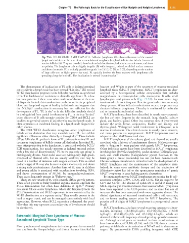
Fig. 73.4 FOLLICULAR LYMPHOMA (FL). Follicular lymphoma (FL) shows effacement of the normal
lymph node architecture because of an accumulation of neoplastic lymphoid follicles that lack the features of
reactive follicles (A). They are crowded, show back-to-back localization, lack distinct mantle zones, and show
no polarity. The lymphoma cells are highly irregular (B) with elongated, twisted, or clefted nuclear contours
and dense chromatin. FL is typically graded into grade 1 or 2 (1/2; C, D), or 3 (E), depending on the number
of large cells seen at higher power (see text). FL typically involves the bone marrow with lymphoma cells
spreading along the bone (F). This localization is termed “paratrabecular.”
The phenomenon of localization of FL cells to isolated germinal Isaacson and Wright as part of the spectrum of mucosa-associated
5
centers within a lymph node has been termed FL in situ. The revised lymphoid tissue (MALT) lymphomas. MALT lymphomas are char-
WHO classification proposes in situ follicular neoplasia as a preferred acterized by a heterogeneous cellular composition that includes
term. The likelihood of evolution to clinically significant FL is low marginal-zone or centrocyte-like cells, monocytoid B cells, small
for these patients, if there is no other evidence of disease at the time lymphocytes, and plasma cells (Fig. 73.5A). In most cases, large
of diagnosis. Indeed, this translocation can be found in the peripheral transformed cells are infrequent. Reactive germinal centers are nearly
blood and lymphoid organs of healthy individuals, and suggests that always present. When follicular colonization occurs, the process may
the BCL2/JH translocation is necessary but not sufficient for the simulate follicular lymphoma. Clonality is confirmed by molecular
development of FL. “FL in situ” or in situ follicular neoplasia should and or immunohistochemical studies.
be distinguished from partial involvement by FL. In the true “in situ” MALT lymphomas have been described in nearly every anatomic
lesion clusters of B cells strongly positive for CD10 and BCL2 are site but are most frequent in the stomach, lung, thyroid, salivary
localized to germinal centers in an otherwise reactive lymph node. It gland, and lacrimal gland. Other less common sites of involvement
often represents an incidental finding, in a lymph node biopsied for include the orbit, breast, conjunctiva, bladder and kidney, and
other reasons. thymus gland. Widespread nodal involvement is infrequent, as is
The 2008 WHO classification recognizes other lymphomas of marrow involvement. The clinical course is usually quite indolent,
follicle center derivation that may resemble nodal FL, but exhibit and many patients are asymptomatic. MALT lymphomas tend to
12
significant differences either clinically or biologically. These include relapse in other MALT-associated sites.
diffuse follicular FL, pediatric forms of FL, primary intestinal FL and MALT lymphomas of the salivary gland and thyroid are usually
cutaneous lymphomas of follicle center cell derivation. Intestinal FL, associated with a history of autoimmune diseases. Helicobacter gas-
most often presenting in the duodenum, is associated with the BCL2/ tritis is frequent in most patients with gastric MALT lymphomas.
IGH translocation, but usually presents as isolated mucosal polyps Other infectious agents have been described in MALT lymphomas
5
with a low risk of dissemination. FL in the pediatric age group is involving skin (Borrelia burgdorferi), ocular adnexae (Chlamydia psit-
histologically diverse. Most nodal cases are cytologically high grade, taci), and small intestine (Campylobacter jejuni); however, in this
composed of blastoid cells, but are usually localized, and may be latter group a causal relationship has not yet been demonstrated.
cured in a number of instances with surgical excision. This so-called Chronic antigen stimulation is critical to both the development of a
pediatric type of FL may also be seen in adults more rarely, and shows MALT lymphoma and the maintenance of the neoplastic state.
a strong male predominance. Another form of FL seen in children Indeed, in some cases antibiotic therapy and the eradication of
and young adults is associated with translocations involving IRF4, Helicobacter pylori has led to the spontaneous remission of gastric
and shows overexpression of MUM1 by immunohistochemistry. MALT lymphoma in cases lacking genetic aberrations.
These cases frequently present in Waldeyer ring. 13 By immunophenotype MALT lymphomas are positive for B-cell–
There are rare variants of FL with a mainly diffuse growth pattern. associated antigens CD19, CD20, and CD22, but are negative for
These often present as bulky localized inguinal masses, and lack the CD5 and CD10. The absence of cyclin D1 is useful in ruling out
12
BCL2 translocation but often have deletions at 1p36. Primary MCL, especially in intestinal disease. Rare cases of MALT lymphoma
cutaneous follicle center lymphoma, which also frequently lacks the have been reported to be CD5-positive, and in some but not all
BCL2 translocation and BCL2 expression, is now considered by the instances this has been associated with more aggressive disease. The
2
WHO classification as a separate entity. They usually present in the clinical significance of increased transformed cells is still uncertain,
head or upper trunk, and can be managed conservatively with local and no formal grading system exists for MALT lymphoma. The
approaches. However, when BCL2 expression is detected, the possi- putative cell of origin of MALT lymphoma is a postgerminal center
bility that this may represent a secondary site of involvement should B-cell.
be considered. MALT lymphomas also have several recurring cytogenetic abnor-
malities, including t(11;18)(q21;q21), t(1;14)(p22;q32), t(14;18)
Extranodal Marginal-Zone Lymphoma of Mucosa- (q32;q21), t(3;14)(q27;q32), and t(3;14)(p14.1;q32), which are
observed with variable frequency, often depending upon the anatomic
Associated Lymphoid Tissue Type site. Although several genes are involved in these translocations, at
least three of them, (t(11;18), t(1;14), and t(14;18), share a common
Most lymphomas of marginal-zone derivation present in extranodal pathway, which leads to the activation of NF-κB and its downstream
sites and have the histopathologic and clinical features identified by targets. By genome–wide DNA profiling integrated with GEP,