
Page 1347 - Hematology_ Basic Principles and Practice ( PDFDrive )
P. 1347
Chapter 73 The Pathologic Basis for the Classification of Non-Hodgkin and Hodgkin Lymphomas 1193
CD10 BCL6
B B C C D D
MUM1 CD138
A A E E F F G G
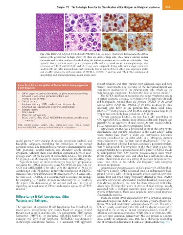
Fig. 73.6 DIFFUSE LARGE B-CELL LYMPHOMA. The low-power illustration demonstrates the diffuse
nature of the process (A). At high power (B), there are sheets of large cells. Those with a vesicular nuclear
chromatin and variable numbers of nucleoli along the nuclear membrane are referred to as centroblasts. These
typically have a germinal center gene expression profile and a germinal center immunophenotype with
expression of CD10 and BCL6 (C and D). Those cases composed of large cells with a single prominent
nucleolus (E) are called immunoblastic and commonly have an activated B-cell (ABC) gene expression profile
and an ABC phenotype with expression of MUM-1, CD138 (F and G), and IFR-4. The correlation of
morphology and immunophenotype is not always exact.
clinical behavior, and often presents with advanced stage and bone
Varied Basis for the Recognition of Diverse Entities Among Aggressive
B-Cell Neoplasms marrow involvement. The relevance of the microenvironment and
recruitment mechanism of the inflammatory cells, which are the
• Cell of origin, in part as determined by gene expression profiling main histologic component, has been the focus of recent studies. 21
Activated B cell versus germinal center B cell The WHO classification recognizes that some lymphomas arising
Thymic B cell of PMBL in certain anatomic sites may have distinctive features both clinically
• Clinical factors and biologically. Among these are primary DLBCL of the central
Anatomic site, e.g., CNS, mediastinum, intravascular nervous system (CNS) and DLBCL of the testis. DLBCLs in these
Advanced age, background of chronic inflammation sanctuary sites differ at the genomic level from usual nodal
• Etiologic factors DLBCL. 22,23 Both primary CNS DLBCL and intraocular large B-cell
EBV, HHV-8
• Molecular pathogenesis lymphomas commonly have mutations in MYD88.
BCL6, C-MYC, ALK, BCL2, MYD88 (translocations, amplification, Primary cutaneous DLBCL, leg type, has a GEP resembling the
mutation) ABC type of DLBCL, presents most often in older adult females, and
generally has an aggressive clinical course. As with nodal DLBCL,
CNS, Central nervous system; EBV, Epstein-Barr virus; HHV-8, human BCL2 expression is an adverse prognostic factor.
herpesvirus-8; PMBL, primary mediastinal large B cell lymphoma;
EBV-positive DLBCL was a provisional entity in the 2008 WHO
24
classification, and was first recognized in the older adult. More
recent studies have shown a wider age distribution. Decreased
immune surveillance in the older adult, or a tolerogenic immune
25
nuclei generally have vesicular chromatin, prominent nucleoli, and microenvironment may facilitate tumor development. The mor-
basophilic cytoplasm, resembling the centroblasts of the normal phologic spectrum is broad, but most cases have a prominent inflam-
germinal center. The immunoblastic variant is characterized by cells matory background. The prognosis in the older adult is poor but
with prominent central nucleoli, and abundant deeply staining younger patients have a good outcome. EBV-positive DLBCL should
cytoplasm. Although there is no absolute correlation between mor- be distinguished from EBV-positive mucocutaneous ulcer, which
phology and GEP, the majority of centroblastic DLBCL falls into the affects mainly cutaneous sites, and often has a self-limited clinical
GCB group, and the majority of immunoblastic into the ABC group. course. These lesions arise in a setting of decreased immune surveil-
Algorithms based on immunophenotype have been proposed as lance, most often in the elderly, and frequently with iatrogenic
surrogates for cDNA microarray using CD10/BCL6 positivity for immune suppression.
GCB and MUM1/IRF-4 for ABC with the addition of BCL2 in Lymphomatoid granulomatosis is an EBV-positive B-cell lymphop-
combination with IPI and may improve the stratification of DLBCL. roliferative disorder (LPD) associated with an inflammatory back-
Because of emerging differences in the treatment of GCB versus ABC ground rich in T cells. The lung is nearly always involved, with skin,
(or non-GCB) DLBCL, it is recommended to include the subtype kidney, liver and brain being frequently affected as well. DLBCL
by integrating GEP and/or immunohistochemistry (IHC) results in associated with chronic inflammation was first described in association
the report with clarification of the method used and the type of with chronic pyothorax, but now has been associated with EBV-
algorithm. As noted, newer GEP methods may be applicable to FFPE driven large B-cell proliferations in diverse clinical settings, usually
material. 18 associated with a confined anatomic space and a background of
chronic inflammation. These cases appear to have a good prognosis
Diffuse Large B-Cell Lymphomas, Other if successfully resected.
There are several LPDs associated with HHV-8/Kaposi sarcoma–
Variants and Subtypes. associated herpesvirus (KSHV). These include primary effusion lym-
phoma (PEL) and multicentric Castleman disease (MCD). The cells of
The spectrum of aggressive B-cell lymphomas has broadened in PEL are usually coinfected with EBV, and the disease is most often
recent years, incorporating new entities based on unique clinical diagnosed in the setting of human immunodeficiency virus (HIV)
features such as age or anatomic site, viral pathogenesis (EBV, human infection and immunosuppression. While pleural or peritoneal effu-
20
herpesvirus [HHV]-8), or distinctive pathologic features. T cell/ sions are most common, extracavitary PEL can present as a tumor
histiocyte–rich large B-cell lymphoma (THRLBCL) has distinctive mass, usually in extranodal sites. PEL has a phenotype resembling
morphologic and clinical features. It is associated with aggressive that of terminally differentiated B-cells, i.e., plasmablastic (Fig. 73.7).