
Page 1359 - Hematology_ Basic Principles and Practice ( PDFDrive )
P. 1359
Chapter 74 Origin of Hodgkin Lymphoma 1205
GC B
cells
Proliferation
Somatic
hypermutation
Advantagous
V gene Plasma
mutations cell
Rescue from Disadvantageous
apoptosis V gene mutations Selection
HRS
precursor
cell
Differentiation
Apoptotic
Further B cells
transforming Transforming Memory
events events B cell
LP
cell
HRS
cell
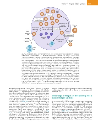
Fig. 74.1 THE GERMINAL CENTER REACTION AND A SCENARIO FOR HODGKIN AND REED-
STERNBERG AND LYMPHOCYTE PREDOMINANT CELL DERIVATION. Antigen-activated B cells
that receive costimulatory signals from T-helper cells establish germinal center (GC) reactions in T-dependent
humoral immune responses. In the GC, the activated B cells undergo massive clonal expansion in the dark
zone. The proliferating GC B cells are called centroblasts. In the centroblasts, the process of somatic hypermuta-
tion is activated, which introduces somatic mutations at a very high rate into rearranged Ig V genes. Centroblasts
migrate into the light zone and become resting centrocytes. Centrocytes are then selected based on their BCR.
Most Ig V gene mutations will be disadvantageous (e.g., when they cause premature stop codons or lead to
amino acid changes that reduce the affinity of the BCR to the immunizing antigen) and will cause apoptotic
death of the respective B cells. GC B cells acquiring affinity-increasing mutations will be positively selected
by interaction with T-helper cells and follicular dendritic cells (not shown). Positively selected centrocytes may
either return to the dark zone for further rounds of proliferation, mutation, and selection, or they differentiate
into memory B cells or plasma cells and exit the GC. LP cells of NLPHL express functional Ig V genes and
proliferate in a follicular microenvironment resembling GC. Thus these cells are likely derived from selected
GC B cells. HRS cells in classical HL often carry destructive Ig V gene mutations, indicating that they derive
from GC B cells that normally would have undergone apoptosis (i.e., preapoptotic GC B cells). Initial
transforming events in HRS cell pathogenesis might have occurred already at a pre–GC B cell differentiation
stage, or they might happen in the GC. In EBV-positive cases, EBV is a good candidate for a factor that allows
GC B cells with destructive Ig V gene mutations to escape from apoptosis.
immunophenotype suggests a B-cell origin. Moreover, LP cells are of typical B cell genes and that their gene expression pattern indicates
located in follicular structures in close association with follicular a relationship to late GC B cells on the way to becoming post-GC
dendritic cells and GC-type T-helper cells, suggesting a close relation- memory B cells. 2
ship with GC B cells. Indeed, LP cells express the transcription factor
BCL6, the main regulator of the GC B cell differentiation program.
They also express activation-induced cytidine deaminase (AID), an Cellular Origin of Hodgkin and Reed-Sternberg Cells in
enzyme that regulates somatic hypermutation and class switching in Classical Hodgkin Lymphoma
GC B cells. Thus all of these phenotypic features point to a GC B
cell origin of LP cells (Table 74.1), and this was further corroborated As mentioned earlier, HRS cells show a peculiar immunophenotype
at the genetic level when clonally rearranged and productive Ig V that does not resemble any normal hematopoietic cell type. HRS cells
region genes were amplified from isolated LP cells. These rearrange- express markers of different cell lineages in a fraction or even most
ments were always somatically mutated. In some cases, intraclonal cases, including B cell genes (e.g., PAX5, IRF4), T cell genes (e.g.,
diversity of V region genes was observed, indicating ongoing somatic NOTCH-1, GATA3), natural killer cell genes (ID2), myeloid genes
hypermutation during clonal expansion. Therefore the Ig V gene (e.g., CD15, CSF1R), and dendritic cell markers (e.g., CCL17,
analysis further strongly supported a GC B cell origin of LP cells (Fig. fascin, restin) (Table 74.1). 1,3,4 Therefore immunohistochemical
74.1). The detection of the same V gene rearrangements in all LP studies could not resolve the cellular origin of HRS cells. In addition,
cells of a given NLPHL case was also important because this firmly the first cell lines established from patients with HL and presumed
established for the first time the monoclonality of the LP cells, a to be derived from HRS cells were either of B-cell origin, T-cell, or
hallmark for tumor cells. A gene expression profiling study of primary nonlymphoid origin (discussed in further detail later). The origin of
LP cells indicated that LP cells have partly downregulated a fraction HRS cells was finally clarified when HRS cells microdissected from