
Page 1354 - Hematology_ Basic Principles and Practice ( PDFDrive )
P. 1354
1200 Part VII Hematologic Malignancies
A A B C C
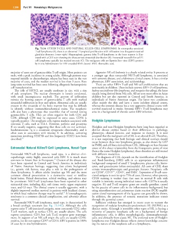
Fig. 73.14 OTHER T-CELL AND NATURAL KILLER-CELL LYMPHOMAS. In enteropathy-associated
T-cell lymphoma (A), there is an abnormal T-lymphoid proliferation with infiltration into the gastrointestinal
glandular elements (center right). Hepatosplenic gamma delta T-cell lymphoma in the bone marrow is illus-
trated with a CD2 stain showing the characteristic sinusoidal distribution (B). Extranodal natural killer cell/T-
cell lymphoma typically has marked necrosis (C). The malignant cells are Epstein-Barr virus (EBV)–positive
by in situ hybridization for EBV encoded RNA (insert). RNA, ribonucleic acid.
majority of cases are of gamma-delta T-cell origin. Most patients are Aggressive NK-cell leukemia is a closely related entity. It presents at
male, with a peak incidence in young adults. Although patients may a younger age than extranodal NK/T-cell lymphoma, is associated
respond initially to chemotherapy, relapse has been seen in the vast with systemic disease, and a fulminant clinical course. It has a similar
majority of cases, and the median survival is less than 3 years. Rare phenotype, EBV association, and epidemiology.
long-term survival has been seen following allogeneic hematopoietic There are other EBV+ T-cell and NK-cell proliferations that are
cell transplantation. seen mainly in children. These include systemic EBV+ T-cell lymphoma,
The cells of HSTCL are usually moderate in size, with a rim hydroa vacciniforme-like lymphoma, and mosquito bite allergy, the latter
of pale cytoplasm. The nuclear chromatin is loosely condensed, usually being derived from NK-cells. All are seen most often in Asian
with small inconspicuous nucleoli. The pattern of infiltration children but are also reported in Central and South America, in
mimics the homing pattern of gamma-delta T cells with marked individuals of Native American origin. The latter two conditions
sinusoidal infiltration in liver and spleen. Abnormal cells are usually affect mainly the skin and have a more indolent clinical course,
present in the sinusoids of the bone marrow but may be difficult whereas the systemic disease has a very aggressive clinical course with
to identify without immunohistochemical stains. The neoplastic survival measured in weeks. Systemic EBV+ T-cell lymphoma may
cells also have a phenotype that resembles that of normal resting arise in a background of chronic active EBV-infection.
gamma-delta T cells. They are often negative for both CD4 and
CD8, although CD8 may be expressed in some cases. CD56 is
typically positive. The neoplastic cells express markers associated with Hodgkin Lymphomas
cytotoxic T cells, such as TIA-1. However, perforin and granzyme
B are usually negative, suggesting that these cells are not activated. Hodgkin and non-Hodgkin lymphoma have long been regarded as
Isochromosome 7q is a consistent cytogenetic abnormality, and is distinct disease entities based on their differences in pathology,
often seen in association with trisomy 8. In addition, activating phenotype, clinical features, and response to therapy. It is now
mutations involving STAT5B and less frequently STAT3 have been accepted that the malignant cell of HL is an altered B cell. Therefore,
described. 36 it is not surprising that both biologic and clinical overlaps should
occur between these two lymphoma groups, as also shown by GEP
in PMBL and cell lines derived from CHL. Although we have become
Extranodal Natural Killer/T-Cell Lymphoma, Nasal-Type aware of this closer relationship from the histogenetic point of view
(hence the name Hodgkin lymphoma), these disorders are still treated
Extranodal NK/T-cell lymphoma, nasal type, is a distinct clini- with different modalities.
copathologic entity highly associated with EBV. It is much more The diagnosis of CHL depends on the identification of Hodgkin
34
common in Asians than in Europeans. Clusters of the disease also and Reed-Sternberg (HRS) cells in an appropriate inflammatory
have been reported in Central and South America in individuals background composed of small T lymphocytes, plasma cells, histio-
of Native American heritage, suggesting that ethnic background cytes, and granulocytes (often eosinophils). All cases of CHL share
i.e., genetic risk factors, may play a role in the pathogenesis of certain immunophenotypic and genotypic features. Neoplastic cells
−
+/−
−
+
these lymphomas. It affects adults (median age 50) and the most are CD30 , CD15 , CD45 , and EMA . Expression of B-cell–asso-
common clinical presentation is a destructive nasal or midline ciated antigens is seen in up to 75% of cases. However, when present,
facial lesion. Palatal destruction, orbital swelling, and edema may CD20 staining is weaker than that seen in normal B cells with
be prominent. NK/T-cell lymphomas have been reported in other variable in intensity among individual tumor cells. CD79a is usually
extranodal sites, including skin, soft tissue, testis, upper respiratory negative. IG and T-cell receptor genes are usually germline, caused
tract, and GI tract. The clinical course is usually aggressive, with a by the paucity of tumor cells in the inflammatory background, but
slightly improved median survival in patients with localized disease, using microdissection and polymerase chain reaction (PCR) amplifi-
37
in which local radiation therapy may be useful. A hemophagocytic cation clonal rearrangement of the Ig genes can generally be shown.
syndrome is a common clinical complication, and adversely affects In addition, the presence of somatic mutations indicates transit
survival. through the germinal center.
Extranodal NK/T-cell lymphoma, nasal type, is characterized by Sufficient evidence has emerged in recent years to warrant the
a broad cytologic spectrum (see Fig. 73.14C). Although the cells recognition of nodular lymphocyte-predominant HL (NLPHL) as a
express some T cell-associated antigens, most commonly CD2, other distinct entity. Although it resembles other types of HL in having a
T-cell markers, such as surface CD3, are usually absent. The cells minority of putative neoplastic cells on a background of benign
express cytoplasmic CD3, but lack T-cell receptor gene rearrange- inflammatory cells, it differs morphologically, immunophenotypi-
ment. In support of an NK-cell origin the cells are usually CD56- cally, and clinically from classic HL. The preferred term of Hodgkin
positive, but do not express CD57 or CD16. EBV is positive in 100% lymphoma over Hodgkin disease reflects current knowledge concern-
of cases by in situ hybridization. ing the nature of the neoplastic cell as a lymphocyte.