
Page 1480 - Hematology_ Basic Principles and Practice ( PDFDrive )
P. 1480
Chapter 82 Diagnosis and Treatment of Diffuse Large B-Cell Lymphoma and Burkitt Lymphoma 1315
Fig. 82.7 BURKITT LYMPHOMA (BL) INVOLVING THE UMBILI-
CUS. This is an 18-year old man who presented with a history of abdominal
pain. Computed tomography scan demonstrated a large intraabdominal mass
extending up to the umbilicus, and a biopsy revealed it to be BL.
children and young adults and is more commonly observed in boys.
Immunodeficiency-associated BL occurs in association with HIV
infection and is approximately 1000 times more common in HIV-
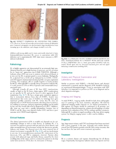
infected individuals. Fig. 82.8 FLUORESCENT IN SITU HYBRIDIZATION SHOWING AN
MYC TRANSLOCATION IN A PATIENT WITH DIFFUSE LARGE
B-CELL LYMPHOMA. This is a break apart probe (LSI-MYC) with one
Pathobiology normal (fused) signal and one abnormal (separated green and red) signal
indicating a translocation involving MYC.
BL is highly aggressive and characterized by an extremely high pro-
liferation fraction and a high fraction of apoptosis, and this accounts
for its “starry sky” appearance (see E-Slide VM03958). Although a Investigation
leukemic phase of BL can occur in patients with advanced disease, it
is very rare for BL to present purely as acute leukemia. Biologically,
+
+
BL is derived from a GCB cell as indicated by its CD20 , CD10 , History and Physical Examination and
and TdT-negative immunohistochemical profile and gene expression Laboratory Investigations
profiling. The neoplastic cells are usually negative or weakly positive
for BCL2. Although EBV is virtually always detected in endemic BL, Similar to patients with DLBCL, a detailed history and physical
it is only present in 25% to 40% of sporadic and immunodeficiency- examination is required, and the diagnosis of BL should be made by
associated cases. an experienced hematopathologist. Given its association with HIV
Although virtually all cases of BL have MYC translocations, infection, it is imperative to perform an HIV test at diagnosis and to
usually at 8q24 to the IG heavy chain region, MYC translocations check hepatitis serologies.
are not specific for BL and can be found in other aggressive B-cell
lymphomas (Fig. 82.7). BL has a unique gene expression signature
that is molecularly distinct from that of DLBCL. Studies have Imaging and Staging
demonstrated that some cases of DLBCL by histology have gene
expression profiles consistent with BL. Given that BL does not As with DLBCL, imaging studies should include chest radiography
respond well to CHOP-based treatments, this distinction by molecu- and CT scanning of the chest, abdomen, and pelvis. The need for
lar profiling is important, and gene expression profiling may be useful additional imaging studies depends on the clinical presentation. A
in rare cases that would otherwise be diagnosed as DLBCL. Addition- BM aspirate and biopsy should be performed in all patients, and
ally, there are cases with a profile intermediate between that of depending on clinical presentation, patients should undergo a lumbar
DLBCL and BL; these cases typically harbor MYC and have a poor puncture with evaluation of the CSF by cytology and flow cytometry.
outcome with CHOP-based regimens. Although BL in adults is staged according to the Ann Arbor staging
system, the Murphy staging system is often used in children.
Clinical Features
Prognosis
The clinical presentation of BL is variable and depends on the epi-
demiologic subtype as well as other factors. In endemic BL, it is Age, large tumor volume, and CNS involvement have been associated
common for patients to present with jaw and other facial disease, and with a poor prognosis in the past. Although early studies demon-
other extranodal sites of involvement include the ileocecum, gonads, strated that HIV-positive patients with BL had a worse outcome, this
kidneys, and breasts. The ileocecal area is the most common site of has not been the case with newer treatment approaches.
disease involvement in sporadic BL (Fig. 82.8), and jaw involvement
is very rare. In immunodeficiency-associated BL, involvement of the
ileocecum, LNs, and BM is commonly observed. Patients often Treatment
present with advanced stage and bulky disease caused by the short
doubling time of the tumor, and it is common for patients to develop BL is a systemic disease and requires chemotherapy for all disease
tumor lysis syndrome (TLS) after the institution of therapy. stages. Importantly, locoregional radiation does not improve survival