
Page 1484 - Hematology_ Basic Principles and Practice ( PDFDrive )
P. 1484
Chapter 83 Virus-Associated Lymphoma 1319
TABLE Viruses and Lymphomagenesis situ hybridization or related techniques, although greatly improved
83.1 in recent years, remain the purview of research laboratories and are
generally not readily applicable to clinical specimens. This reflects the
Virus Viral Genome in Tumor Cell Lymphoma Type relatively low copy number of the viral genome in tumor cells, typi-
EBV Episomal B, T, NK cally 1–200 copies per cell. In contrast, in situ hybridization for the
EBV-encoded ribonucleic acids (RNAs) has emerged as a clinical
KSHV Episomal B 12
laboratory standard. These RNAs are polymerase 3 transcripts that
HTLV-1 Integrated T are expressed at very high copy number (perhaps millions of copies
HIV-1 Absent B per cell) in latently infected cells. The functions of these RNAs are
disputed, but their use for the detection of virus in a variety of tissue
HCV Uncertain B
specimens is generally accepted.
EBV, Epstein-Barr virus; HCV, hepatitis C virus; HIV-1, human Viral antigens are detected by immunohistochemistry. In clinical
immunodeficiency virus type 1; HTLV-1, human T-lymphotropic virus-1; KSHV, laboratories, immunohistochemistry for LMP1 is commonly
Kaposi sarcoma–associated herpesvirus; NK, natural killer.
employed and is sensitive for the detection of EBV in Hodgkin
lymphoma (HL). In a variety of other EBV-associated B- and T-cell
malignancies, expression of LMP1 is more variable. Thus failure to
TABLE Patterns of Epstein-Barr Virus Gene Expression in detect LMP1 expression does not exclude the presence of EBV, except
83.2 Latency perhaps in HL. In principle, detection of EBNA1 should be univer-
EBNA2, EBNA3A, sally applicable, although the low level of antigen expression and the
Latency EBNA1 EBNA3B, EBNA3C LMP1 LMP2A cross-reactivity of available monoclonal antibodies have prevented
immunohistochemistry for this antigen from emerging as a standard
I + tool.
II + + +
III + + + +
EBNA1, Epstein-Barr virus nuclear antigen 1; EBV, Epstein-Barr virus; LMP1, Association With Particular Types of Lymphoma
latent membrane protein 1.
Some lymphoma types are nearly 100% EBV associated, including
endemic BL, extranodal natural killer (NK)/T-cell lymphoma of the
nasal type, early PTLD, lymphomatoid granulomatosis, diffuse large
LMP2
B-cell lymphoma (DLBCL) associated with chronic inflammation,
EBV-positive DLBCL of older adults, and AIDS primary central
LMP1 nervous system (CNS) lymphoma (PCNSL). 13–16 Other lymphoma
B-cell receptor types are variably EBV associated. These include classic HL, PTLDs
occurring many months or years after transplantation, and systemic
AIDS-related lymphoma.
Some lymphoma types appear never or almost never to be EBV
NF-κB associated, including most indolent B-cell lymphomas, although
there is growing evidence that exceptions do exist, particularly in the
Cell setting of immunocompromise. Nodular lymphocyte predominant
survival HL was historically thought to always be EBV negative, but a recent
pathologic review of over 300 cases suggests that 3%–5% are EBV
EBNA1
17
positive. Low-grade lymphomas such as EBV-positive mucosa-
associated lymphoid tissue (MALT) lymphomas have been reported
18
in patients with congenital immunodeficiencies. Whereas low-grade
lymphomas that arise posttransplant are not considered to be PTLD
in the current classification, it is interesting to note that cases of
Transcription EBV-positive, indolent B-cell lymphomas, particularly MALT lym-
factors:
EBNA-LP phomas with a predilection to involve the subcutaneous or soft
19,20
tissues, have been reported in transplant patients.
Thus the
EBNA2
EBNA3A, EBNA3B, spectrum of EBV-associated lymphomas continues to expand, and
EBNA3C
the presence of EBV might be an indication to consider an underlying
immune defect in the patient.
Other lymphomas are typically not EBV associated but have the
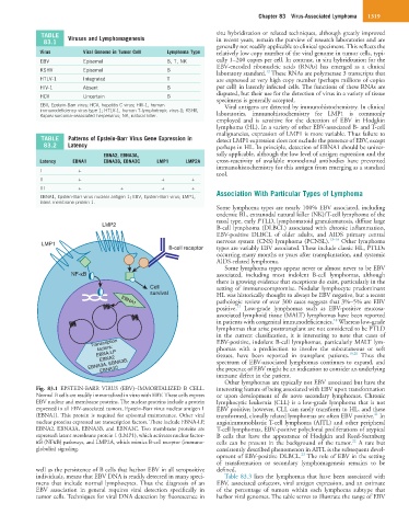
Fig. 83.1 EPSTEIN-BARR VIRUS (EBV)–IMMORTALIZED B CELL. interesting feature of being associated with EBV upon transformation
Normal B cells are readily immortalized in vitro with EBV. These cells express or upon development of de novo secondary lymphomas. Chronic
EBV nuclear and membrane proteins. The nuclear proteins include a protein lymphocytic leukemia (CLL) is a low-grade lymphoma that is not
expressed in all EBV-associated tumors, Epstein-Barr virus nuclear antigen 1 EBV positive; however, CLL can rarely transform to HL, and these
21
(EBNA1). This protein is required for episomal maintenance. Other viral transformed, clonally related lymphomas are often EBV positive. In
nuclear proteins expressed are transcription factors. These include EBNA-LP, angioimmunoblastic T-cell lymphoma (AITL) and other peripheral
EBNA2, EBNA3A, EBNA3B, and EBNA3C. Two membrane proteins are T-cell lymphomas, EBV-positive polyclonal proliferations of atypical
expressed: latent membrane protein 1 (LMP1), which activates nuclear factor- B cells that have the appearance of Hodgkin and Reed-Sternberg
22
κB (NFκB) pathways, and LMP2A, which mimics B-cell receptor (immuno- cells can be present in the background of the tumor. A rare but
globulin) signaling. consistently described phenomenon in AITL is the subsequent devel-
23
opment of EBV-positive DLBCL. The role of EBV in the setting
of transformation or secondary lymphomagenesis remains to be
well as the persistence of B cells that harbor EBV in all seropositive defined.
individuals, means that EBV DNA is readily detected in many speci- Table 83.3 lists the lymphomas that have been associated with
mens that include normal lymphocytes. Thus the diagnosis of an EBV, associated cofactors, viral antigen expression, and an estimate
EBV association in general requires viral detection specifically in of the percentage of tumors within each lymphoma subtype that
tumor cells. Techniques for viral DNA detection by fluorescence in harbor viral genomes. The table serves to illustrate the range of EBV