
Page 1632 - Hematology_ Basic Principles and Practice ( PDFDrive )
P. 1632
Chapter 89 Clinical Approach to Infections in the Compromised Host 1453
TABLE Pulmonary Infiltrates and Their Association With
89.3 Specific Infectious and Noninfectious Etiologies
Radiologic Sign Differential Diagnosis
Interstitial Pulmonary edema
infiltrates Diffuse alveolar damage
Idiopathic pneumonia syndrome
Respiratory viruses: respiratory syncytial virus,
parainfluenza, influenza, adenovirus, enterovirus
Herpes viruses: cytomegalovirus, herpes simplex
virus, varicella zoster virus, human herpes virus
type 6
Pneumocystis pneumonia A
Focal airspace Bacterial pneumonia
disease Fungal pneumonia
Nodules Fungal pneumonia (aspergillosis)
Nocardia
Legionella
Septic bacterial emboli
Mycobacterial infection (with cavitation)
Epstein-Barr virus lymphoproliferative disorder
Relapsed malignancy
Pulmonary embolism (pleural based)
Halo sign or air Aspergillosis
crescent sign
Two common anaerobic infections occur during neutropenia B
at sites where biopsy is difficult or contraindicated. Neutropenic
enterocolitis, also known as typhlitis, manifests as fever, abdominal
pain, and tenderness. CT scan of the abdomen shows signs of
16
right-sided colonic and ileal inflammation. Excessive soft-tissue
swelling of the neck during mucositis can present as a Ludwig angina
variant. Broadly active antianaerobic, aerobic, and possibly antifungal
antimicrobial agents should be added for either of these clinical
findings.
Pulmonary Infiltrates
Pulmonary infections are common in the immunocompromised host
(see box on Approach to Pulmonary Infiltrates). Plain chest radiog-
raphy is a good initial screen but lacks the sensitivity of CT, which
generally provides more useful information in terms of characterizing
the nature of an infiltrate and assists the pulmonologist in determin- C
ing where to direct the bronchoscope for highest yield. A specific
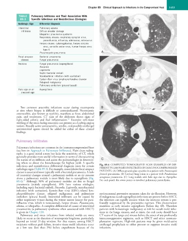
infectious and noninfectious differential diagnosis exists for certain Fig. 89.4 COMPUTED TOMOGRAPHY SCAN EXAMPLES OF DIF-
radiologic signs (Table 89.3). For example, consolidative focal airspace FERENT PULMONARY INFILTRATES IN IMMUNOCOMPROMISED
disease is associated most typically with a bacterial pneumonia. A halo PATIENTS. (A) Diffuse ground-glass opacities in a patient with Pneumocystis
of interstitial changes around a pulmonary nodule or an air crescent jirovecii pneumonia. (B) Cavitary lung lesion in a patient with Pseudomonas
above a pulmonary nodule is most likely due to aspergillosis (Fig. aeruginosa pneumonia. (C) Lung nodule with halo sign due to Aspergillus.
17
89.4). Although 90% of pulmonary nodules are due to fungal For each panel, the arrow points to involved pulmonary parenchyma.
pneumonia (mainly aspergillosis), 10% have various etiologies,
including septic bacterial emboli, Nocardia, Legionella, mycobacterial
infection (with cavitation), Epstein-Barr virus (EBV)–related lym-
phoproliferative disease, relapsed malignancy, and pulmonary environmental preventive measures taken for air filtration. However,
embolism (pleural based). Interstitial infiltrates can be caused by if endogenous occult aspergillosis infections are present before HSCT,
either respiratory viruses during the winter season (except for para- the infection can rapidly escalate when the immune system is pro-
influenza virus, which is nonseasonal), herpes viruses, Pneumocystis, foundly suppressed by the preparative regimen. This phenomenon
edema, or idiopathic. A complete differential of causes of interstitial manifests as early invasive aspergillosis (before day 40). Therefore
pneumonitis (a pulmonary syndrome often associated with HSCT) patients with hematologic malignancy at risk for occult mold infec-
is given in Chapter 110. tions in the lungs, sinuses, and at times, the oral cavity should have
Pulmonary and sinus infections from inhaled molds are more CT scans of the lungs and sinuses before the onset of any profoundly
likely to occur as the duration of neutropenia lengthens, particularly immunosuppressive regimens, such as HSCT and select nontrans-
beyond an initial 21-day window. For that reason, among HSCT plantation regimens. High-risk patients may be given mold-active
recipients without graft failure, invasive tissue mold infections occur antifungal prophylaxis to either prevent or suppress invasive mold
at a low rate (less than 3%) before engraftment because of the infections.