
Page 2212 - Hematology_ Basic Principles and Practice ( PDFDrive )
P. 2212
Chapter 132 Thrombocytopenia Caused by Platelet Destruction, Hypersplenism, or Hemodilution 1959
The anatomy of the spleen is uniquely suited for its function; Physiologic Platelet Sequestration
progressive branching of the splenic artery into trabecular and central
arteries helps separate the plasma from the cellular elements (see Radiolabeled platelet studies have shown that approximately 30% of
Chapter 160). The central arteries arise perpendicularly from the the total platelet mass exists as a freely exchangeable pool in the
trabecular arteries and skim the plasma layer from the cells. Soluble spleen. Because the normal platelet life span is 9–10 days, platelets
antigens in the plasma are delivered to the white pulp, where phago- spend approximately one-third of their lives, or 3 days, within the
cytic cells process them and antibody production is initiated. spleen. In patients with hypersplenism, up to 90% of the platelets
A cell-rich, hemoconcentrated fraction of the blood is delivered can be found in the spleen.
to the red pulp. Some of this blood flows directly to the splenic veins After labeled platelets are injected, there is accumulation in both
(the closed system), but most moves into the splenic cords (the open the liver and the spleen. An initial, irreversible phase of hepatic
system). Here, the cellular elements percolate through a meshwork uptake occurs. This equilibrates during the first 5 minutes and may
of reticulum fibers, reticuloendothelial cells, and supporting cells to reflect hepatic clearance of platelets damaged during the labeling
reach the splenic sinuses. The cells enter the sinuses by passing procedure. Simultaneously, there is a slow increase in activity over the
through narrow fenestrations in the basement membrane of the spleen that peaks in about 20 minutes. Splenic platelet uptake is thus
endothelial cells lining the sinuses. The blood exits through the dependent on input (spleen blood flow) and output (clearance).
splenic vein into the portal system. Because the veins in the portal The splenic platelet pool size can be decreased and the platelet
system lack valves, any increase in portal pressure is transmitted to count increased with intravenous infusions of epinephrine in normal
the splenic microcirculation. persons and in patients with splenomegaly. By contrast, isoprenaline
The spleen plays a number of important roles. It is the largest increases the splenic pool size. Splenic blood flow increases with
lymphoid organ in the body and contributes to host defense by increasing spleen size, although perfusion (flow per unit of tissue
clearing microorganisms and antibody-coated cells. The spleen is also volume) falls. Blood flow can be increased in some inflammatory
important for antibody synthesis, especially antibodies directed disorders (e.g., SLE) without an increase in spleen size. A marked
against soluble antigens. The filtering function of the spleen includes increase or decrease in splenic perfusion alters the proportion of
(1) culling (removal of damaged or senescent cells and bacteria), (2) platelets within the spleen.
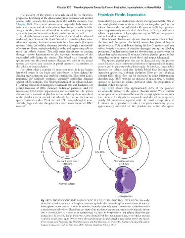
pitting (removal of RBC inclusion bodies or parasites), and (3) Fig. 132.3 shows why approximately 30% of the platelets
1
remodeling (reticulocyte sequestration and maturation). The spleen are normally present in the spleen. Because about 5% of cardiac
also serves as a reservoir of platelets (accommodating about one-third output goes to the spleen and because the average splenic transit time
of the platelet mass in normal individuals). By contrast, the human (i.e., the time for the platelet to pass through the spleen) is approxi-
spleen contains less than 2% of the total RBC mass, although in some mately 10 minutes—compared with the usual average time of
animals (dogs and cats), the spleen is a much more important RBC 1 minute for a platelet to make a complete circulatory pass—
reservoir. approximately one-third of the platelets are within the spleen
95%
5% 5%-25%
~1 min
Normal transit time Normal transit time
(~10 min) (~10 min)
Normal spleen
Hypersplenism
Fig. 132.3 PHYSIOLOGIC AND PATHOLOGIC PLATELET SPLENIC SEQUESTRATION. Normally,
about 5% of cardiac output is to the spleen; however, a platelet that enters the spleen spends about 10 minutes
there (splenic transit time = 10 min). In contrast, it usually takes only about 1 minute for a platelet to make
a circulatory pass elsewhere. Thus about one-third of the platelets at any one time are located within the spleen:
(5% × 10 min):(95% × 1 min), or an approximate 1 : 2 ratio. In hypersplenism, the splenic blood flow can
increase by a factor of 5, that is, from 5% to 25% of total blood flow per minute. Thus, even without increase
in splenic transit time, up to 70% or more of the platelets can be exchangeably sequestered within the spleen.
(From Arnold DM, Warkentin TE: Thrombocytopenia and thrombocytosis. In: Wilson WC, Grande CM, Hoyt DB, editors:
Trauma: Critical care, vol. 2, New York, 2007, Informa Healthcare USA, p 983.)