
Page 2563 - Hematology_ Basic Principles and Practice ( PDFDrive )
P. 2563
2286 Part XIII Consultative Hematology
I J K L
M N O P
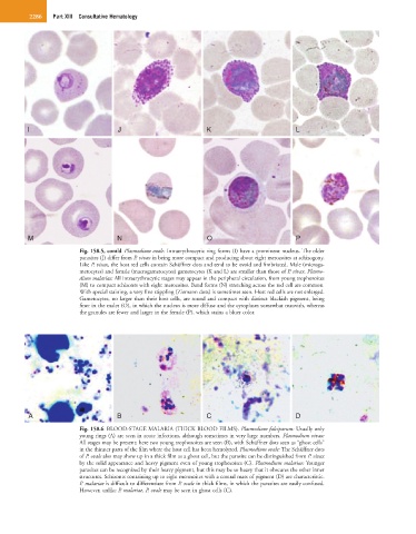
Fig. 158.5, cont’d Plasmodium ovale: Intraerythrocytic ring forms (I) have a prominent nucleus. The older
parasites (J) differ from P. vivax in being more compact and producing about eight merozoites at schizogony.
Like P. vivax, the host red cells contain Schüffner dots and tend to be ovoid and fimbriated. Male (microga-
metocytes) and female (macrogametocytes) gametocytes (K and L) are smaller than those of P. vivax. Plasmo-
dium malariae: All intraerythrocytic stages may appear in the peripheral circulation, from young trophozoites
(M) to compact schizonts with eight merozoites. Band forms (N) stretching across the red cell are common.
With special staining, a very fine stippling (Ziemann dots) is sometimes seen. Host red cells are not enlarged.
Gametocytes, no larger than their host cells, are round and compact with distinct blackish pigment, being
finer in the males (O), in which the nucleus is more diffuse and the cytoplasm somewhat mauvish, whereas
the granules are fewer and larger in the female (P), which stains a bluer color.
A B C D
Fig. 158.6 BLOOD-STAGE MALARIA (THICK BLOOD FILMS). Plasmodium falciparum: Usually only
young rings (A) are seen in acute infections, although sometimes in very large numbers. Plasmodium vivax:
All stages may be present; here two young trophozoites are seen (B), with Schüffner dots seen as “ghost cells”
in the thinner parts of the film where the host cell has been hemolyzed. Plasmodium ovale: The Schüffner dots
of P. ovale also may show up in a thick film as a ghost cell, but the parasite can be distinguished from P. vivax
by the solid appearance and heavy pigment even of young trophozoites (C). Plasmodium malariae: Younger
parasites can be recognized by their heavy pigment, but this may be so heavy that it obscures the other inner
structures. Schizonts containing up to eight merozoites with a central mass of pigment (D) are characteristic.
P. malariae is difficult to differentiate from P. ovale in thick films, in which the parasites are easily confused.
However, unlike P. malariae, P. ovale may be seen in ghost cells (C).