
Page 546 - Hematology_ Basic Principles and Practice ( PDFDrive )
P. 546
Chapter 34 Approach to Anemia in the Adult and Child 461
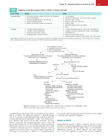
TABLE Comparison of the More Common Causes of Anemia in Children and Adult
34.3
Type of Anemia Children Adults
Hypoproliferative • Nutritional deficiency (most commonly iron deficiency) • Iron deficiency
• Acute inflammation • Anemia of inflammation (anemia of chronic disease)
• Transient erythroblastopenia of childhood • Anemia of renal disease
• Acquired aplastic anemia • Folate or vitamin B 12 deficiency
• Marrow replacement caused by malignancy • Drugs or toxins
• Pure RBC aplasia (viral or idiopathic)
• MDS
Hemolytic • Inherited hemoglobinopathies • Inherited hemoglobinopathies with milder manifestations
• Inherited membrane disorders • Inherited membrane disorders with milder manifestations
• Autoimmune hemolytic anemia • G6PD deficiency
• Microangiopathic hemolytic anemia • Autoimmune hemolytic anemia
• Microangiopathic hemolytic anemia (DIC, TTP, HUS, aHUS)
aHUS, Atypical hemolytic uremic syndrome; DIC, disseminated intravascular coagulation; G6PD, glucose-6-phosphate dehydrogenase; HUS, hemolytic uremic syndrome;
MDS, myelodysplastic syndrome; RBC, red blood cell; TTP, thrombotic thrombocytopenic purpura.
For a newborn review:
1. Complete blood cell count
2. Reticulocyte count
3. Peripheral blood smear
Reticulocyte count Reticulocyte count
Corrected reticulocyte count <2% or absolute Corrected reticulocyte count >2% or absolute
reticulocyte count <100,000/uL reticulocyte count ≥100,000/uL
Congenital hypoplastic anemia Direct antiglobulin test
Transcobalamin II deficiency
Negative Positive
Immune hemolytic anemia
ABO
MCV Rh
Minor blood group
Low Normal or elevated
Chronic intrauterine blood loss
α-Thalassemia syndrome Review of peripheral blood smear
Normal Abnormal
Blood loss Membrane disorders
Iatrogenic (blood sampling) Hereditary spherocytosis
Fetomaternal/fetoplacental Hereditary elliptocytosis
Twin-to-twin Hereditary stomatocytosis
Internal hemorrhage Metabolic disorders
Galactosemia G6PD deficiency
Infection Pyruvate kinase deficiency
Bacterial Infection
Viral Disseminated intravascular
Toxoplasmosis coagulation
Congenital syphilis
Rare causes
Hexokinase deficiency
Fig. 34.3 APPROACH TO THE DIFFERENTIAL DIAGNOSIS OF ANEMIA IN A NEWBORN. G6PD,
Glucose-6-phosphate dehydrogenase; MCV, mean corpuscular volume.
hyperbilirubinemia that occurs. After true hemolysis has been identi- Alternatively, ethnic background and family history may be helpful
fied in an infant, the differential diagnosis is relatively limited in arriving at the appropriate diagnosis.
(Fig. 34.3). Immune-mediated hemolysis may result from ABO, Rh,
17
or minor blood group incompatibility. Other causes include meta-
bolic disorders and disorders of the RBC membrane. Of note, Anemia in Adults
however, is the fact that hemoglobinopathies, such as sickle cell
disease and β-thalassemia, are silent during the newborn period and Hypoproliferative anemia in adults is relatively common. If acute
only become manifest at 4 to 6 months of age when the fetal-to-adult blood loss is excluded, hypoproliferative causes are the most common
hemoglobin transition has been completed. Newborn screening entities associated with anemia in adults. These are iron deficiency,
programs in the United States may provide salient information in inflammation (anemia of chronic disease), and renal disease
this regard on the presence or absence of a hemoglobinopathy. (Fig. 34.4). The megaloblastic anemias that represent maturation