
Page 547 - Hematology_ Basic Principles and Practice ( PDFDrive )
P. 547
462 Part V Red Blood Cells
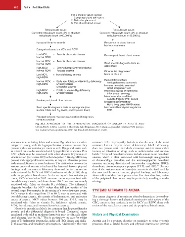
For a child or adult review:
1. Complete blood cell count
2. Reticulocyte count
3. Peripheral blood smear
Reticulocyte count Reticulocyte count
Corrected reticulocyte count <2% or absolute Corrected reticulocyte count >2% or absolute
reticulocyte count <100,000/uL reticulocyte count ≥100,000/uL
Hypoproliferative anemia Response to blood loss or
hemolytic anemia
Categorize based on MCV and RDW
Low MCV, = Anemia of chronic disease Review peripheral blood smear
Normal RDW
Normal MCV, = Anemia of chronic disease
Normal RDW Send specific diagnostic tests as
appropriate
High MCV, = Chemotherapy/antivirals/alcohol
Normal RDW Aplastic anemia Differential diagnoses/
Low MCV, = Iron deficiency anemia tests to obtain:
High RDW
Hemoglobinopathies/
Normal MCV, = Early iron, folate, or vitamin B 12 deficiency hemoglobin electrophoresis
High RDW Myelodysplasia Immune hemolytic anemias/
Dimorphic anemia direct antiglobulin test
High MCV, = Folate or vitamin B 12 deficiency Infectious causes of hemolysis/
High RDW Myelodysplasia thick smear, serology
Membrane abnormalities/
Review peripheral blood smear osmotic fragility; PNH screen
Metabolic abnormalities/
Heinz body prep; G6PD assay
Send specific diagnostic tests as appropriate (iron Mechanical hemolysis/coagulation
studies, folate and B levels, erythropoietin level) tests
12
Proceed to bone marrow examination if diagnosis
remains unclear
Fig. 34.4 APPROACH TO THE DIFFERENTIAL DIAGNOSES OF ANEMIA IN ADULTS AND
CHILDREN. G6PD, Glucose-6-phosphate dehydrogenase; MCV, mean corpuscular volume; PNH, paroxys-
mal nocturnal hemoglobinuria; RDW, red blood cell distribution width.
abnormalities, including folate and vitamin B 12 deficiency, are often common RBC enzymopathy (which is also the one of the most
categorized along with the hypoproliferative anemias because they common human enzyme defect deficiencies), G6PD deficiency,
present with a low reticulocyte count as well. Drugs and toxins such does not present until individuals encounter oxidant stress either
as ethanol can also be associated with hypoproliferative anemia. Pure because of infection or drugs such as sulfonamides and antima-
20
RBC aplasia may be associated with other diseases (thymoma) or larials. Acquired hemolytic anemias include autoimmune hemolytic
18
viral infection (parvovirus B19) or be idiopathic. Finally, MDS may anemia, which is often associated with hematologic malignancies
present with hypoproliferative anemia, as may an infiltrative process or rheumatologic disorders, and the microangiopathic hemolytic
such as myelofibrosis or acute leukemia. The distinction between the anemias, including disseminated intravascular coagulation (DIC),
21
various causes of anemia is facilitated by historical factors, physical thrombotic thrombocytopenic purpura (TTP), and HUS. Distinc-
findings, and concomitant laboratory abnormalities in conjunction tion of the various causes of hemolytic anemia is also facilitated by
with review of the MCV and RBC distribution width (RDW) along the associated historical features, physical findings, and laboratory
with the peripheral blood smear. In the setting of a low reticulocyte abnormalities of the clinical presentation. For these disorders, review
count, MCV values below 70 fL are most commonly associated with of the peripheral blood smear may be particularly revealing as to the
iron-deficiency anemia, and those above 120 fL are most commonly etiology.
associated with folate or vitamin B 12 deficiency. The differential
diagnosis broadens for MCV values that fall just outside of the
normal range. For example, in the setting of a low reticulocyte count, SYSTEMIC APPROACH TO ANEMIA
MCV values in the range from 75 to 80 fL may be associated with
iron-deficiency anemia, the anemia of inflammation, and endocrine The correct diagnosis of anemia can often be determined by combin-
causes of anemia. MCV values between 100 and 110 fL may be ing a thorough history and physical examination with review of the
associated with folate or vitamin B 12 deficiency, aplastic anemia, CBC, concentrating particularly on the MCV and RDW, along with
MDS, liver disease, and immune hemolytic anemias. review of the reticulocyte count and the peripheral blood smear.
Hemolytic anemia in adults is less common than hypoproliferative
anemia, and the differential diagnosis is broad. Congenital causes
associated with mild to moderate hemolysis may be clinically silent History and Physical Examination
19
until detected later in life. This is particularly the case for milder
cases of β-thalassemia intermedia, sickle cell (SC) disease and sickle- Anemia can be a primary disorder or secondary to other systemic
+
β -thalassemia, and hereditary spherocytosis. Additionally, the most processes, thus a careful history and physical examination provide