
Page 551 - Hematology_ Basic Principles and Practice ( PDFDrive )
P. 551
466 Part V Red Blood Cells
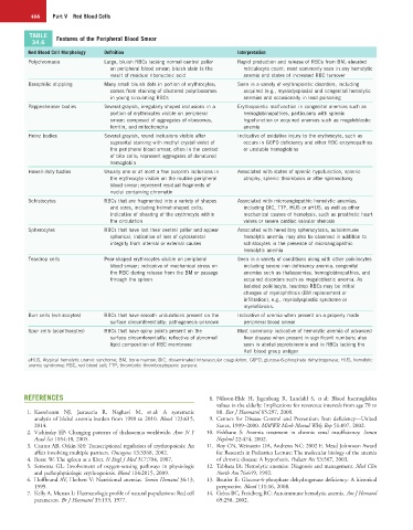
TABLE Features of the Peripheral Blood Smear
34.6
Red Blood Cell Morphology Definition Interpretation
Polychromasia Large, bluish RBCs lacking normal central pallor Rapid production and release of RBCs from BM; elevated
on peripheral blood smear; bluish stain is the reticulocyte count; most commonly seen in any hemolytic
result of residual ribonucleic acid anemia and states of increased RBC turnover
Basophilic stippling Many small bluish dots in portion of erythrocytes; Seen in a variety of erythropoietic disorders, including
comes from staining of clustered polyribosomes acquired (e.g., myelodysplasia) and congenital hemolytic
in young circulating RBCs anemias and occasionally in lead poisoning
Pappenheimer bodies Several grayish, irregularly shaped inclusions in a Erythropoietic malfunction in congenital anemias such as
portion of erythrocytes visible on peripheral hemoglobinopathies, particularly with splenic
smear; composed of aggregates of ribosomes, hypofunction or acquired anemias such as megaloblastic
ferritin, and mitochondria anemia
Heinz bodies Several grayish, round inclusions visible after Indicative of oxidative injury to the erythrocyte, such as
supravital staining with methyl crystal violet of occurs in G6PD deficiency and other RBC enzymopathies
the peripheral blood smear, often in the context or unstable hemoglobins
of bite cells; represent aggregates of denatured
hemoglobin
Howell-Jolly bodies Usually one or at most a few purplish inclusions in Associated with states of splenic hypofunction, splenic
the erythrocyte visible on the routine peripheral atrophy, splenic thrombosis or after splenectomy
blood smear; represent residual fragments of
nuclei containing chromatin
Schistocytes RBCs that are fragmented into a variety of shapes Associated with microangiopathic hemolytic anemias,
and sizes, including helmet-shaped cells; including DIC, TTP, HUS or aHUS, as well as other
indicative of shearing of the erythrocyte within mechanical causes of hemolysis, such as prosthetic heart
the circulation valves or severe cardiac valvular stenosis
Spherocytes RBCs that have lost their central pallor and appear Associated with hereditary spherocytosis, autoimmune
spherical; indicative of loss of cytoskeletal hemolytic anemia; may also be observed in addition to
integrity from internal or external causes schistocytes in the presence of microangiopathic
hemolytic anemia
Teardrop cells Pear-shaped erythrocytes visible on peripheral Seen in a variety of conditions along with other poikilocytes
blood smear; indicative of mechanical stress on including severe iron deficiency anemia, congenital
the RBC during release from the BM or passage anemias such as thalassemias, hemoglobinopathies, and
through the spleen acquired disorders such as megaloblastic anemia. As
isolated poikilocyte, teardrop RBCs may be initial
changes of myelophthisis (BM replacement or
infiltration), e.g., myelodysplastic syndrome or
myelofibrosis.
Burr cells (echinocytes) RBCs that have smooth undulations present on the Indicative of uremia when present on a properly made
surface circumferentially; pathogenesis unknown peripheral blood smear
Spur cells (acanthocytes) RBCs that have spiny points present on the Most commonly indicative of hemolytic anemia of advanced
surface circumferentially; reflective of abnormal liver disease when present in significant numbers; also
lipid composition of RBC membrane seen in abetalipoproteinemia and in RBCs lacking the
Kell blood group antigen
aHUS, Atypical hemolytic uremic syndrome; BM, bone marrow; DIC, disseminated intravascular coagulation; G6PD, glucose-6-phosphate dehydrogenase; HUS, hemolytic
uremic syndrome; RBC, red blood cell; TTP, thrombotic thrombocytopenic purpura.
REFERENCES 8. Nilsson-Ehle H, Jagenburg R, Landahl S, et al: Blood haemoglobin
values in the elderly: Implications for reverence intervals from age 70 to
1. Kassebaum NJ, Jasrasaria R, Naghavi M, et al: A systematic 88. Eur J Haematol 65:297, 2000.
analysis of blobal anemia burden from 1990 to 2010. Blood 123:615, 9. Centers for Disease Control and Prevention: Iron deficiency—United
2014. States, 1999–2000. MMWR Morb Mortal Wkly Rep 51:897, 2002.
2. Vichinsky EP: Changing patterns of thalassemia worldwide. Ann N Y 10. Fishbane S: Anemia treatment in chronic renal insufficiency. Semin
Acad Sci 1054:18, 2005. Nephrol 22:474, 2002.
3. Cantor AB, Orkin SH: Transcriptional regulation of erythropoiesis: An 11. Roy CN, Weinstein DA, Andrews NC: 2002 E. Mead Johnnson Award
affair involving multiple partners. Oncogene 13:3368, 2002. for Research in Pediatrics Lecture: The molecular biology of the anemia
4. Rosse W: The spleen as a filter. N Engl J Med 317:704, 1987. of chronic disease: A hypothesis. Pediatr Res 53:507, 2003.
5. Semenza GL: Involvement of oxygen-sensing pathways in physiologic 12. Tabbara IA: Hemolytic anemias: Diagnosis and management. Med Clin
and pathophysiologic erythropoiesis. Blood 114:2015, 2009. North Am 76:649, 1992.
6. Hoffbrand AV, Herbert V: Nutritional anemias. Semin Hematol 36:13, 13. Beutler E: Glucose-6-phosphate dehydrogenase deficiency: A historical
1999. perspective. Blood 111:16, 2008.
7. Kelly A, Munan L: Haematologic profile of natural populations: Red cell 14. Gehrs BC, Freidberg RC: Autoimmune hemolytic anemia. Am J Hematol
parameters. Br J Haematol 35:153, 1977. 69:258, 2002.