
Page 550 - Hematology_ Basic Principles and Practice ( PDFDrive )
P. 550
Chapter 34 Approach to Anemia in the Adult and Child 465
A B C D E
F G H I J
K L M N O
P Q R S T
U V W X Y
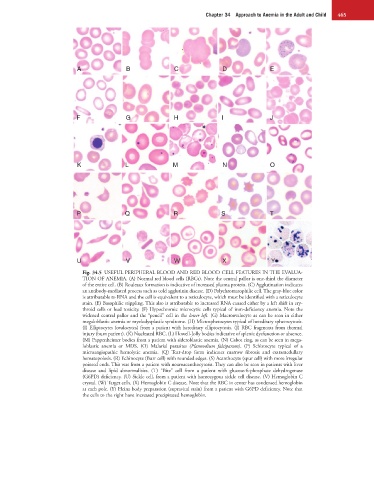
Fig. 34.5 USEFUL PERIPHERAL BLOOD AND RED BLOOD CELL FEATURES IN THE EVALUA-
TION OF ANEMIA. (A) Normal red blood cells (RBCs). Note the central pallor is one-third the diameter
of the entire cell. (B) Rouleaux formation is indicative of increased plasma protein. (C) Agglutination indicates
an antibody-mediated process such as cold agglutinin disease. (D) Polychromatophilic cell. The gray-blue color
is attributable to RNA and the cell is equivalent to a reticulocyte, which must be identified with a reticulocyte
stain. (E) Basophilic stippling. This also is attributable to increased RNA caused either by a left shift in ery-
throid cells or lead toxicity. (F) Hypochromic microcytic cells typical of iron-deficiency anemia. Note the
widened central pallor and the “pencil” cell in the lower left. (G) Macroovalocyte as can be seen in either
megaloblastic anemia or myelodysplastic syndrome. (H) Microspherocytes typical of hereditary spherocytosis.
(I) Elliptocytes (ovalocytes) from a patient with hereditary elliptocytosis. (J) RBC fragments from thermal
injury (burn patient). (K) Nucleated RBC. (L) Howell-Jolly bodies indicative of splenic dysfunction or absence.
(M) Pappenheimer bodies from a patient with sideroblastic anemia. (N) Cabot ring, as can be seen in mega-
loblastic anemia or MDS. (O) Malarial parasites (Plasmodium falciparum). (P) Schistocyte typical of a
microangiopathic hemolytic anemia. (Q) Tear-drop form indicates marrow fibrosis and extramedullary
hematopoiesis. (R) Echinocyte (Burr cell) with rounded edges. (S) Acanthocyte (spur cell) with more irregular
pointed ends. This was from a patient with neuroacanthocytosis. They can also be seen in patients with liver
disease and lipid abnormalities. (T) “Bite” cell from a patient with glucose-6-phosphate dehydrogenase
(G6PD) deficiency. (U) Sickle cell, from a patient with homozygous sickle cell disease. (V) Hemoglobin C
crystal. (W) Target cells. (X) Hemoglobin C disease. Note that the RBC in center has condensed hemoglobin
at each pole. (Y) Heinz body preparation (supravital stain) from a patient with G6PD deficiency. Note that
the cells to the right have increased precipitated hemoglobin.