
Page 548 - Hematology_ Basic Principles and Practice ( PDFDrive )
P. 548
Chapter 34 Approach to Anemia in the Adult and Child 463
valuable insight into the potential cause. Fatigue often accompanies can accompany or even predate the anemia associated with vitamin
anemia, but it is very nonspecific and may be related to systemic B 12 deficiency, findings such as loss of vibration or position sense in
illness. Nonetheless, determining the concomitant presence of a the extremities may be relevant. 24
systemic inflammatory disorder, infection, or malignancy that may
be associated with fatigue can be critical in determining the underly-
ing causes of anemia in both adults and children. The medical history Reticulocyte Count
may also be quite informative. For example, a history of diabetes
mellitus can be associated with significantly impaired renal produc- As a marker of RBC production, the reticulocyte count provides
tion of erythropoietin even in the setting of only a mildly elevated essential information in directing the initial investigation of anemia.
creatinine level. Because certain medications may be associated with Modern flow cytometers accurately determine the reticulocyte count
BM depression or, alternatively, the development of autoimmune using fluorescent probes that bind to the residual ribonucleic acid
25
hemolytic anemia, all pharmacologic agents, prescribed and over the present in newly released RBCs. These measurements are useful,
counter, including alternative medicines, should be reviewed. Occu- accurate, and reflect the state of erythropoiesis. However, when sig-
pational history is occasionally relevant, as in the case of individuals, nificant numbers of nucleated RBCs or nuclear debris are present in
such as welders, who might have been exposed to lead or other the peripheral blood, this diagnostic accuracy declines, and manual
potentially BM toxic agents. Social history can be important. A counting methods are generally preferable.
history of intravenous drug use might suggest the possibility of virally When the reticulocyte count is reported as a percentage, it needs
transmitted diseases, such as HIV, which may be associated with to be adjusted for the total number of RBCs present. This correction
anemia. Dietary history is also very important, particularly in young can be made by multiplying the reticulocyte count by the patient’s
and elderly individuals with anemia. The finding of pica in adults hematocrit divided by an age- and sex-appropriate normal hematocrit.
(most commonly ice chips or cornstarch) is well known to be associ- No such correction is necessary when the reticulocyte count is
22
ated with iron-deficiency anemia. Ingestion of paint chips may reported as an absolute number or when it is converted to an absolute
suggest the need to investigate the possibility of toxic lead ingestion. number by multiplying the percentage by the RBC number (in
A family history of anemia is highly relevant in the evaluation of RBC/µL).
children with anemia. However, it is also relevant in adults because In the absence of anemia, the normal absolute reticulocyte count
+
certain congenital anemias, such as milder forms of sickle β thalas- is between 25,000 and 75,000/µL. In the presence of anemia, an
semia and hereditary spherocytosis, occasionally first become clini- absolute reticulocyte count of less than 75,000/µL is indicative of a
cally apparent in adulthood. hypoproliferative process, and an absolute reticulocyte count of
The significance of pallor on physical examination is in many ways greater than 100,000/µL is indicative of hemolysis or an appropriate
similar to the historic feature of fatigue: it is a common but nonspe- erythropoietic response to blood loss (see Table 34.2). Reticulocyte
cific finding. More specific findings may be found in certain types of counts between 75,000 and 100,000/µL require interpretation in the
anemia. For example, angular cheilitis (cracking at the edges of the context of other available clinical data, including the severity of
lips) and koilonychia (spooning of the nails) may accompany iron- anemia present.
deficiency anemia. Splenomegaly may be present in patients with
anemia arising from a wide variety of different causes. When present
early in life, it is suggestive of a congenital hemolytic anemia, such Mean Corpuscular Volume and Red Blood Cell
as thalassemia, sickle cell disease, or hereditary spherocytosis. When Distribution Width From the Complete Blood Count
found for the first time later in life, splenomegaly may indicate an
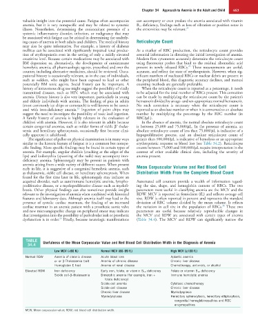
acquired disorder, such as autoimmune hemolytic anemia, lympho- Automated cell counters provide a wealth of information regard-
proliferative disease, or a myeloproliferative disease such as myelofi- ing the size, shape, and hemoglobin content of RBCs. The two
brosis. Other physical findings can also sometimes provide insight parameters most useful in classifying anemia are the MCV and the
relevant to the investigation of anemia when combined with historical RDW. MCV is reported in femtoliters (fL) and reflects average cell
features and laboratory data. Although anemia itself may lead to the size. RDW is often reported in percent and represents the standard
presence of systolic cardiac murmurs, the finding of an increased deviation of RBC volume divided by the mean volume. It reflects
26
cardiac murmur in an anemic patient with a prosthetic aortic valve the variation in cell size in the population of RBCs. These two
and new microangiopathic change on peripheral smear may indicate parameters are useful because relatively reproducible changes in
that investigation into the possibility of perivalvular leak or prosthetic the MCV and RDW are associated with certain types of anemia
23
dysfunction is in order. Finally, because neurologic manifestations (Table 34.4). The MCV and RDW can significantly narrow the
TABLE Usefulness of the Mean Corpuscular Value and Red Blood Cell Distribution Width in the Diagnosis of Anemia
34.4
Low MCV (<80 fL) Normal MCV (80–99 fL) High MCV (≥100 fL)
Normal RDW Anemia of chronic disease Acute blood loss Aplastic anemia
α- or β-Thalassemia trait Anemia of chronic disease Chronic liver disease
Hemoglobin E trait Anemia of renal disease Chemotherapy, antivirals, or alcohol
Elevated RDW Iron deficiency Early iron, folate, or vitamin B 12 deficiency Folate or vitamin B 12 deficiency
Sickle cell-β–thalassemia Dimorphic anemia (for example, iron + Immune hemolytic anemia
folate deficiency)
Sickle cell anemia Cytotoxic chemotherapy
Sickle cell disease Chronic liver disease
Chronic liver disease Myelodysplasia
Myelodysplasia Hereditary spherocytosis, hereditary elliptocytosis,
congenital hemoglobinopathies and RBC
enzymopathies
MCW, Mean corpuscular value; RDW, red blood cell distribution width.