
Page 549 - Hematology_ Basic Principles and Practice ( PDFDrive )
P. 549
464 Part V Red Blood Cells
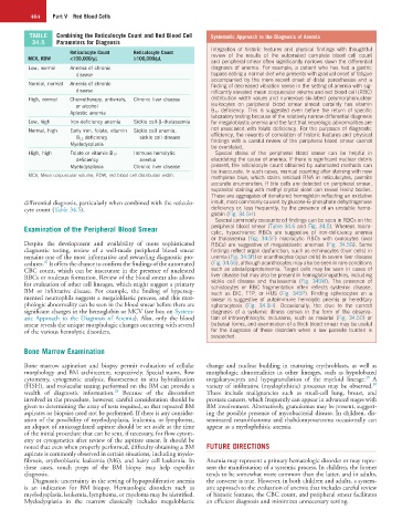
TABLE Combining the Reticulocyte Count and Red Blood Cell Systematic Approach to the Diagnosis of Anemia
34.5 Parameters for Diagnosis
Integration of historic features and physical findings with thoughtful
Reticulocyte Count Reticulocyte Count review of the results of the automated complete blood cell count
MCV, RDW <100,000/µL ≥100,000/µL
and peripheral smear often significantly narrows down the differential
Low, normal Anemia of chronic diagnosis of anemia. For example, a patient who has had a gastric
disease bypass eating a normal diet who presents with gradual onset of fatigue
accompanied by the more recent onset of distal paresthesias and a
Normal, normal Anemia of chronic finding of decreased vibration sense in the setting of anemia with sig-
disease nificantly elevated mean corpuscular volume and red blood cell (RBC)
High, normal Chemotherapy, antivirals, Chronic liver disease distribution width values and numerous six-lobed polymorphonuclear
or alcohol leukocytes on peripheral blood smear almost certainly has vitamin
Aplastic anemia B 12 deficiency. This is suggested even before the return of specific
laboratory testing because of the relatively narrow differential diagnosis
Low, high Iron-deficiency anemia Sickle cell-β–thalassemia for megaloblastic anemia and the fact that neurologic abnormalities are
Normal, high Early iron, folate, vitamin Sickle cell anemia, not associated with folate deficiency. For the purposes of diagnostic
B 12 deficiency sickle cell disease efficiency, the rewards of correlation of historic features and physical
Myelodysplasia findings with a careful review of the peripheral blood smear cannot
be overstated.
High, high Folate or vitamin B 12 Immune hemolytic Special stains of the peripheral blood smear can be helpful in
deficiency anemia elucidating the cause of anemia. If there is significant nuclear debris
Myelodysplasia Chronic liver disease present, the reticulocyte count obtained by automated methods can
be inaccurate. In such cases, manual counting after staining with new
MCV, Mean corpuscular volume; RDW, red blood cell distribution width. methylene blue, which stains residual RNA in reticulocytes, permits
accurate enumeration. If bite cells are detected on peripheral smear,
supravital staining with methyl crystal violet can reveal Heinz bodies.
These are aggregates of denatured hemoglobin reflecting an oxidative
differential diagnosis, particularly when combined with the reticulo- insult, most commonly caused by glucose-6-phosphate dehydrogenase
cyte count (Table 34.5). deficiency or, less frequently, by the presence of an unstable hemo-
globin (Fig. 34.5H).
Several commonly encountered findings can be seen in RBCs on the
Examination of the Peripheral Blood Smear peripheral blood smear (Table 34.6 and Fig. 34.5). Whereas micro-
cytic, hypochromic RBCs are suggestive of iron-deficiency anemia
or thalassemia (Fig. 34.5F) macrocytic RBCs with ovalocytes (oval
Despite the development and availability of more sophisticated RBCs) are suggestive of megaloblastic anemias (Fig. 34.5G). Some
diagnostic testing, review of a well-made peripheral blood smear findings reflect organ dysfunction, such as echinocytes (burr cells) in
remains one of the most informative and rewarding diagnostic pro- uremia (Fig. 34.5R) or acanthocytes (spur cells) in severe liver disease
27
cedures. It offers the chance to confirm the findings of the automated (Fig. 34.5S), although acanthocytes may also be seen in rare conditions
CBC count, which can be inaccurate in the presence of nucleated such as abetalipoproteinemia. Target cells may be seen in cases of
RBCs or rouleaux formation. Review of the blood smear also allows liver disease but may also be present in hemoglobinopathies, including
for evaluation of other cell lineages, which might suggest a primary sickle cell disease and thalassemia (Fig. 345W). The presence of
schistocytes or RBC fragmentation often reflects systemic disease,
BM or infiltrative disease. For example, the finding of hyperseg- such as DIC, TTP, or HUS (Fig. 345P). Finding spherocytes on a
mented neutrophils suggests a megaloblastic process, and this mor- smear is suggestive of autoimmune hemolytic anemia or hereditary
phologic abnormality can be seen in the blood smear before there are spherocytosis (Fig. 34.5H). Occasionally, the clue to the correct
significant changes in the hemoglobin or MCV (see box on System- diagnosis of a systemic illness comes in the form of the observa-
atic Approach to the Diagnosis of Anemia). Also, only the blood tion of intraerythrocytic inclusions, such as malarial (Fig. 34.5O) or
smear reveals the unique morphologic changes occurring with several babesial forms, and examination of a thick blood smear may be useful
of the various hemolytic disorders. for the diagnosis of these disorders when a low parasite burden is
suspected.
Bone Marrow Examination
Bone marrow aspiration and biopsy permit evaluation of cellular change and nuclear budding in maturing erythroblasts, as well as
morphology and BM architecture, respectively. Special stains, flow morphologic abnormalities in other lineages, such as hypolobated
29
cytometry, cytogenetic analysis, fluorescence in situ hybridization megakaryocytes and hypogranulation of the myeloid lineage. A
30
(FISH), and molecular testing performed on the BM can provide a variety of infiltrative (myelophthisic) processes may be observed.
28
wealth of diagnostic information. Because of the discomfort These include malignancies such as small-cell lung, breast, and
involved in the procedure, however, careful consideration should be prostate cancers, which frequently can appear in advanced stages with
given to determining the array of tests required, so that repeated BM BM involvement. Alternatively, granulomas may be present, suggest-
aspirates or biopsies need not be performed. If there is any consider- ing the possible presence of mycobacterial disease. In children, dis-
ation of the possibility of myelodysplasia, leukemia, or lymphoma, seminated neuroblastoma and rhabdomyosarcoma occasionally can
an aliquot of anticoagulated aspirate should be set aside at the time appear as a myelophthisic anemia.
of the initial procedure that can be sent, if necessary, for flow cytom-
etry or cytogenetics after review of the aspirate smear. It should be
noted that even when properly performed, difficulty obtaining a BM FUTURE DIRECTIONS
aspirate is commonly observed in certain situations, including myelo-
fibrosis, erythroblastic leukemia (M6), and hairy cell leukemia. In Anemia may represent a primary hematologic disorder or may repre-
these cases, touch preps of the BM biopsy may help expedite sent the manifestation of a systemic process. In children, the former
diagnosis. tends to be somewhat more common than the latter, and in adults,
Diagnostic uncertainty in the setting of hypoproliferative anemia the converse is true. However, in both children and adults, a system-
is an indication for BM biopsy. Hematologic disorders such as atic approach to the evaluation of anemia that includes careful review
myelodysplasia, leukemia, lymphoma, or myeloma may be identified. of historic features, the CBC count, and peripheral smear facilitates
Myelodysplasia in the marrow classically includes megaloblastic an efficient diagnosis and minimizes unnecessary testing.