
Page 554 - Hematology_ Basic Principles and Practice ( PDFDrive )
P. 554
Chapter 35 Pathophysiology of Iron Homeostasis 469
Reticuloendothelial of iron delivery during its lifetime in the circulation. Apotransferrin
macrophages is a true carrier that is not lost in delivering iron; the half-life of the
Muscle, other protein is about 8 days. In an iron-replete 70-kg man, the amount
parenchymal cells of transferrin-bound iron in the plasma at any given time is only
about 3 mg, but more than 30 mg of iron moves through this
transport compartment each day (see Fig. 35.1). Most (approximately
Circulating GI 24 mg Fe/d) of this iron is used for erythropoiesis.
red blood Tf tract Transferrin receptors on the cell surface selectively bind monofer-
cells ric or diferric transferrin. Two different isoforms of the transferrin
receptor exist, encoded by two separate genes. The two glycoproteins
have similar extracellular structures but distinct roles in iron homeo-
stasis. Transferrin receptor 1 is ubiquitously expressed and functions
Erythroid Hepatocytes as the physiologic transferrin iron importer on all iron-requiring cells.
marrow Transferrin receptor 2 is expressed only in hepatocytes, functioning
in the control of iron supply by regulating hepcidin expression (see
later), and in erythroid precursors, coordinating erythropoiesis with
Functional iron iron availability (see later). Transferrin receptor 1 is a transmem-
11
Macrophage storage iron brane glycoprotein dimer composed of two identical subunits linked
Hepatocyte storage iron by a disulfide bond. Each transferrin receptor 1 can bind two mol-
Transport iron ecules of transferrin; if each transferrin is diferric, the dimeric receptor
Sites of hepcidin control can carry a total of four atoms of transferrin-bound iron. The affinity
of iron entry into plasma of transferrin receptor 1 for transferrin depends both on the iron
content of transferrin and on the pH. With amounts of iron-bearing
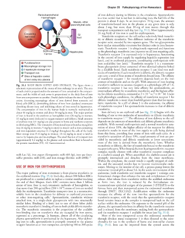
Fig. 35.1 BODY IRON SUPPLY AND STORAGE. The figure shows a transferrin sufficient to saturate receptors at a physiologic pH of 7.4,
schematic representation of the routes of iron exchange in an adult. The area transferrin receptor 1 has very little affinity for apotransferrin; an
of each circle is proportional to the amount of iron contained in the compart- intermediate affinity for monoferric transferrin; and the highest affin-
−9
−9
ment, and the width of each arrow is proportional to the daily flow of iron ity for diferric transferrin, estimated at 2 × 10 to 7 × 10 M. Under
from one compartment to another. Double slashes indicate the sites of hepcidin such physiologic conditions, the affinity of transferrin receptor 1 for
action, decreasing macrophage release of iron derived from senescent red diferric transferrin is more than fourfold greater than that for mono-
blood cells (RBCs), diminishing delivery of iron from duodenal enterocytes ferric transferrin. At a pH of about 5 in the endosome, the affinity
absorbing dietary iron, and inhibiting release of iron stored in hepatocytes. of transferrin receptor 1 for apotransferrin increases to that of diferric
The concentration of iron in the human body is normally maintained at transferrin.
about 40 mg/kg in women and about 50 mg/kg in men. The major portion Iron delivery to an erythroid cell (see Fig. 35.4) begins with the
of iron is found in the erythron as hemoglobin iron (28 mg/kg in women; binding of one or two molecules of monoferric or diferric transferrin
4,5
32 mg/kg in men) dedicated to oxygen transport and delivery. Small amounts to transferrin receptor 1. The efficiency of iron delivery to the cell
of erythron iron (<1 mg/kg) are also present in heme and nonheme enzymes depends on the amounts of monoferric and diferric plasma transferrin
in developing RBCs. The remainder of functional iron is found as myoglobin available. With normal erythropoiesis and a normal transferrin satu-
iron (4 mg/kg in women; 5 mg/kg in men) in muscle and as iron-containing ration of about 33%, the higher affinity of the receptor for diferric
and iron-dependent enzymes (1–2 mg/kg) throughout the cells of the body. transferrin results in most of the iron supply to cells being derived
Most storage iron (5–6 mg/kg in women; 10–12 mg/kg in men) is held in from this form, providing four atoms of iron with each cycle. At a
reserve by hepatocytes and macrophages. The small fraction of transport iron transferrin saturation of about 19%, equal amounts of iron are pro-
(approximately 0.2 mg/kg) in the plasma and extracellular fluid is bound to vided by monoferric and diferric transferrin; at lower saturations,
the protein transferrin (Tf). GI, Gastrointestinal. most of the iron is derived from the monoferric form. Whether
monoferric or diferric, the fate of transferrin bound to the transferrin
receptor is the same. When bound, the iron-bearing transferrin−receptor
complex rapidly clusters with other transferrin−receptor complexes
miR-Let-7d), iron export (ferroportin; miR-485-3p), iron use (iron- in a clathrin-coated pit. When assembled, the clathrin-coated pit is
sulfur proteins: miR-210), and iron storage (ferritin: miR-200b). 10 promptly internalized and detaches from the inner membrane.
Within the cytoplasm, the coated vesicle is rapidly stripped of clath-
rin, and the uncoated vesicles fuse to become multivesicular endo-
USE OF IRON FOR ERYTHROPOIESIS somes. Moving to the interior of the cell, a proton pump lowers the
endosome internal pH to about 5.6. In the acidic environment of the
The major pathway of iron movement is from plasma transferrin to endosome, both transferrin and transferrin receptor 1 undergo con-
the erythroid marrow (Fig. 35.4). Each day, almost 200 billion RBCs formational changes that enhance the rate and completeness of iron
are produced in a normal adult to replace a similar number reaching release. After release from transferrin within the acidified endosome
the end of their lifespan. Each RBC contains more than 1 billion as ferric iron, the iron is reduced by the ferrireductase six-
atoms of iron, four in each tetrameric molecule of hemoglobin, so transmembrane epithelial antigen of the prostate 3 (STEAP3) to the
18
that more than 200 quintillion (200 × 10 ) atoms of iron are needed ferrous form and then transported across the endosomal membrane
daily for erythropoiesis. Transferrin transports iron in a nonreactive, through DMT1 (SLC11A2). Acidification within the endosome
soluble form in the circulation for delivery to erythroid precursors increases the affinity of the now iron-free apotransferrin for the
5
or other iron-requiring cells. Apotransferrin, transferrin without transferrin receptor, with the result that the apotransferrin−receptor
attached iron, is a single-chain glycoprotein with two structurally bond remains intact as the complex is transported back to the cell
similar lobes. Binding of a ferric ion to one of these lobes yields surface within the endosome. On exposure to the neutral pH of the
monoferric transferrin; binding of ions to both yields diferric transfer- plasma, the apotransferrin loses its affinity for the transferrin receptor
rin. The transferrin saturation is the proportion of the available and is released from the membrane, making both the apotransferrin
iron-binding sites on transferrin that are occupied by iron atoms, and the transferrin receptor 1 available for reuse (see Fig. 35.4).
expressed as a percentage. In humans, almost all of the circulating Most of the iron transported across the endosomal membrane
plasma apotransferrin is synthesized by the hepatocyte. After deliver- through divalent metal transporter 1 is then directed to the mito-
ing iron to cells, apotransferrin is promptly returned to the plasma chondria for use in the synthesis of heme and iron-sulfur clusters
to again function as an iron transporter, completing 100 to 200 cycles (Fig. 35.4). Iron can be imported from the cytosol across the