
Page 922 - Hematology_ Basic Principles and Practice ( PDFDrive )
P. 922
Chapter 56 Conventional and Molecular Cytogenomic Basis of Hematologic Malignancies 805
(10%–20%), and potential for high cure rate (>80%) if appropriate
treatment, based on its genetic profile, is initiated. Initial workup may
include conventional karyotyping, FISH studies, RT-PCR, and anti-
PML antibodies. Longitudinal monitoring of disease with Q-PCR is
currently recommended to provide early intervention if relapse
should occur.
Less than 1% of patients with AML have the Ph chromosome (see
A Fig. 56.26, bottom left) and it remains a controversial entity as it is
not listed in the WHO 2016 classification as a separate entity. Ph +
AML may be cytogenetically distinguished from the myeloid blast
crisis of CML by monosomy 7 (also frequently found in Ph + ALL)
and inv(16). Moreover, NPM1 mutations found in 40% to 60% of
AML as well as in Ph + AML have not been described in Ph-positive
CML. All Ph + AML examined to date showed a unique loss of Ig
genes with a specific genomic signature, suggesting a distinct biologic
entity. Both P210 BCR-ABL1 and P190 BCR-ABL1 were observed, but P190
appears to be prevalent. Before TKI therapy, the median survival of
the Ph chromosome +AML was 9 months and has been improved to
a median survival of 18 months (range 6–71) with the use of imatinib
therapy. In rare patients with AML, late appearance of a Ph chromo-
B some either as a sole abnormality or in a clone showing t(8;21) is
taken as evidence that the Ph chromosome in these patients is a
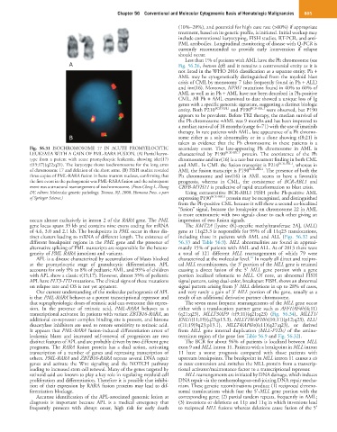
Fig. 56.31 ISOCHROMOSOME 17 IN ACUTE PROMYELOCYTIC secondary event. The late-appearing Ph chromosome in AML is
LEUKEMIA WITH A GAIN OF PML-RARA FUSION. (A) Partial karyo- characterized by P190 BCR-ABL1 protein. The coexistence of the Ph
type from a patient with acute promyelocytic leukemia, showing ider(17) chromosome and inv(16) is a rare but recurrent finding in both CML
t(15;17)(q22;q21). The karyotype shows isochromosome for the long arms and AML. In CML the fusion transcript is P210 BCR-ABL1 , whereas in
of chromosome 17 and deletion of the short arms. (B) FISH studies revealed AML the fusion transcript is P190 BCR-ABL1 . The presence of both the
three copies of PML-RARA fusion in bone marrow nucleus, confirming that Ph chromosome and inv(16) in AML seems to have a favorable
the first event in the pathogenesis was PML-RARA fusion and the subsequent prognosis, whereas in CML, the coexistence of BCR-ABL1 and
event was a structural rearrangement of isochromosome. (From Cheng L, Zhang CBFB-MYH11 is predictive of rapid transformation to blast crisis.
DY, editors: Molecular genetic pathology, Totowa, NJ, 2008, Humana Press, a part Using extrasensitive BCR-ABL1 FISH probe Ph-positive AML
of Springer Science.) expressing P190 BCR-ABL1 protein may be recognized, and distinguished
from the Ph-positive CML because it will show a second co-localized
“fusion” signal, because the breakpoint on chromosome 22 in AML
is more centromeric with two signals closer to each other giving an
occurs almost exclusively in intron 2 of the RARA gene. The PML impression of two fusion signals.
gene locus spans 35 kb and contains nine exons coding for mRNA The KMT2A [lysine (K)-specific methyltransferase 2A], (MLL)
of 4.6, 3.0 and 2.1 kb. The breakpoints in PML occur in three dis- gene at 11q23.3 is responsible for 95% of all 11q23 translocations,
tinct clusters leading to mRNA of different length. The existence of including those in patients with AML and ALL (Figs. 56.32 and
different breakpoint regions in the PML gene and the presence of 56.33 and Table 56.9). MLL abnormalities are found in approxi-
alternative splicing of PML transcripts are responsible for the hetero- mately 15% of patients with AML and ALL. As of 2013 there were
geneity of PML RARA junctions and variants. a total of 121 different MLL rearrangements of which 79 were
16
APL is a disease characterized by accumulation of blasts blocked characterized at the molecular level. In nearly all direct and recipro-
at the promyelocytic stage of granulocytic differentiation. APL cal MLL recombinomes, the 3′ portion of the MLL gene is retained
accounts for only 5% to 8% of pediatric AML, and 95% of children causing a direct fusion of the 5′ MLL gene portion with a gene
with APL show a classic t(15;17). However, almost 35% of pediatric portion localized telomeric to MLL. Of note, an abnormal FISH
APL have FLT3-ITD mutations. The clinical signs of these mutations signal pattern, using dual-color, breakapart FISH, shows an abnormal
on relapse rate and OS is not yet apparent. signal pattern arising from 3′ MLL deletions in up to 28% of cases,
Our current understanding of the molecular pathogenesis of APL and very rarely a gain of 3′ MLL portion of the gene, usually as a
is that PML-RARA behaves as a potent transcriptional repressor and result of an additional derivative partner chromosome.
that supraphysiologic doses of retinoic acid can overcome this repres- The seven most frequent rearrangements of the MLL gene occur
sion. In the presence of retinoic acid, PML-RARA behaves as a either with a translocation partner gene such as AFF1/AF4/t(4;11)
transcriptional activator. In patients with variant ZBTB16-RARA, an (q21;q23), MLLT3/AF9 /t(9;11)(q21;q23) (Fig. 56.34), MLLT1/
additional co-repressor complex binding site is present, and histone ENL/t(11;19)(q23;p13.3), MLLT10/AF10/t(10;11)(p12;q23), ELL/
deacetylase inhibitors are used to restore sensitivity to retinoic acid. t(11;19)9q23;p13.1), MLLT4/AF6/t(6;11)(q27;q23), or derived
It appears that PML-RARA fusion-induced differentiation arrest of from MLL gene internal duplication (MLL-PTDs) of the amino-
leukemic blasts and increased self-renewal of progenitors are two terminus region of the gene (see Table 56.9 and Fig. 56.32).
distinct features of APL and are probably driven by two different gene The BCR for about 96% of patients is localized between MLL
programs. The RARA fusion protein has a dual action, activating exon 9 and MLL intron 11. Patients with a breakpoint in MLL intron
transcription of a number of genes and repressing transcription of 11 have a worse prognosis compared with those patients with
others. PML-RARA and ZBTB16-RARA repress several DNA repair upstream breakpoints. The breakpoint in MLL intron 11 causes a cis
genes and activate the Wnt signaling and the NOTCH pathway to trans conversion and switches the MLL protein from a transcrip-
leading to increased stem cell renewal. Many of the genes targeted by tional activator/maintenance factor to a transcriptional repressor.
retinoid acid are known to play a key role in regulating myeloid cell MLL rearrangements are initiated by DNA damage, which induces
proliferation and differentiation. Therefore it is possible that inhibi- DNA repair via the nonhomologous-end-joining DNA repair mecha-
tion of their expression by RARA fusion proteins may lead to dif- nism. These genetic recombinations produce (1) reciprocal chromo-
ferentiation blockage. somal translocations which fuse the 5′-MLL gene portion with the
Accurate identification of the APL-associated genomic lesion at corresponding gene; (2) partial tandem repeats, frequently in AML;
diagnosis is important because APL is a medical emergency that (3) inversions or deletions on 11p and 11q in which inversions lead
frequently presents with abrupt onset, high risk for early death to reciprocal MLL fusions whereas deletions cause fusion of the 5′