
Page 1103 - Williams Hematology ( PDFDrive )
P. 1103
1078 Part VIII: Monocytes and Macrophages Chapter 68: Production, Distribution, and Activation of Monocytes and Macrophages 1079
GM-CSF has a broader myeloid target profile. It is produced by surface glycoprotein interactions with a selected substratum; T-helper
many cells, including macrophages themselves, especially after inflam- type 2 (Th2) cytokines, such as IL-4 and IL-13, act through a common
34
matory stimuli such as lipopolysaccharide (LPS), and it enhances pro- receptor chain and signaling pathway to enhance macrophage fusion.
duction of monocytes and macrophages with a different morphology DNA synthesis, a feature of high-turnover granulomata associated
to that induced by M-CSF. GM-CSF is required for myeloid DC differ- with infection, can result in abortive cell division and cell death. These
entiation in vitro and has been widely used, alone and in combination macrophage-derived giant cells are distinct from syncytia induced by
with cytokines such as IL-4 or transforming growth factor (TGF)-β, to fusogenic virus infection, especially paramyxoviruses and retroviruses
produce DC from mouse marrow or from human monocytes in cell such as the human immunodeficiency virus.
culture. 31,32 Its targeted deletion in mice or genetic loss of function
mutants of its specific receptor chain in humans results in pulmonary
alveolar proteinosis, associated with defective alveolar macrophage HETEROGENEITY
metabolism of pulmonary surfactant. 33 Monocytes are defined as the population of differentiated cells present
in the circulation, with classical morphologic features (Chap. 67), and
Survival, Differentiation, and Turnover Overview include the less-well defined precursors able to give rise to myeloid DCs
Once the cells have acquired the characteristics of mature monocytes/ and osteoclasts. Because of their ready availability from human blood
macrophages, they display considerable heterogeneity in morphology and the sensitive methods now available to analyze their phenotype
and phenotypic plasticity. In general, their proliferative potential is lim- ex vivo (FACS, microarray, immunochemistry, and cytochemistry),
ited, and their life span can vary from less than 1 day to many months, human monocytes have been more amenable to study, whereas in the
depending on their microenvironment, infections, and other stimuli. mouse, analysis of precursor–product relationship and tissue distribution
Although terminally differentiated, macrophages remain extremely have provided new insights into the fate and heterogeneity of the circu-
active in messenger RNA and protein synthesis, with complex, often lating population. The number of monocytes in the circulation depends
characteristic profiles of gene expression, depending on innate and on constitutive, steady-state production and delivery from marrow, pos-
acquired immune stimuli and cellular interactions. Tissue macrophages sibly from marginated pools in spleen, as well as adhesion and diapedesis
are relatively resistant to apoptosis, compared with neutrophils, but this in response to unknown stimuli and enhanced recruitment in response
feature changes during infection. Their active membrane turnover and to peripheral stimuli such as infection and inflammation. M-CSF and
endocytosis make them susceptible to toxic agents, making them targets glucocorticoids affect their level and phenotype, as do metabolic stim-
for clearance by surviving macrophages. Sublethal injury and infection uli; Chap. 70 describes clinical conditions that give rise to monocytosis.
can also induce autophagy, increasingly recognized as an important The biochemical properties and functions of monocytes are described in
component of inflammatory and infectious diseases. Chap. 67. They are relatively radioresistant once entering the circulation,
The remarkable ability of macrophages to undergo homotypic where they persist for 12 to 48 hours as motile cells, with an ability to
cell–cell fusion results in giant cell formation. This is a feature of osteo- engulf particles and to adhere transiently or more stably to arterial as well
clast differentiation, depending on M-CSF and the tumor necrosis as microvascular endothelium, thus modulating their phagocytic ability.
factor (TNF) family member receptor activator of nuclear factor-κB Depending on interactions with the vessel wall and local differentiation,
ligand (RANKL), which act on monocytic precursors to yield catabolic monocytes are able to crawl along and patrol the intravascular surface
20
cells able to excavate and remodel living bone. Local adhesion and ruf- utilizing CD11a, a β -integrin–dependent property. Mature macro-
2
fling of their plasma membrane are associated with focal, polarized phages lining the endothelium can also detach and recirculate, for exam-
release of H and hydrolytic enzymes by monocyte-derived osteoclasts. ple, filled with lipid stores as foam cells in atherosclerosis, and circulate
+
The attempted uptake of non- or poorly degradable foreign materials heavily laden with erythroid breakdown products in malaria.
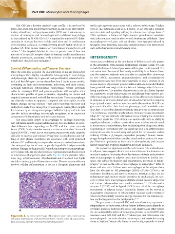
induces “foreign-body giant cells,” with distinct properties; macrophage- The presence of significant numbers of immune cells and molecules
derived giant cells are also characteristic of granulomatous diseases such in adipose tissue suggest vibrant interactions between the immune and
as tuberculosis (Langerhans giant cells; Fig. 68–3) and parasitic infec- metabolic systems. In obesity, the inflammatory infiltrate and activation
tions (e.g., schistosomiasis). Mycobacterial and ill-defined host lipids state of macrophages in adipose tissue may contribute to insulin resis-
are able to induce giant cell formation in vitro. The mechanism of fusion tance. The cellular localization and inflammatory potentials of macro-
36
35
involves cellular differentiation to induce a fusogenic phenotype and phages, as well as the ratio of macrophages to adipocytes, differ in
obese and lean mice. In lean mice, macrophages in the adipose tissue
have the alternate or M2 phenotype (ARG1+CD206+CD301+), are
uniformly distributed, and serve a protective function as they are less
inflammatory and promote insulin sensitivity by producing IL-10; how-
ever, in obese mice, macrophages distribute around necrotic adipocytes,
and induce inflammation and insulin resistance. 35,37 CC-chemokine
receptor 2 (CCR2) and its ligand (CCL2) are critical for macrophage
38
recruitment to adipose tissue. Metabolic disease can be viewed as
maladaptive consequence of inflammation-induced insulin resistance,
which may beneficially conserve energy resources for the immune sys-
tem combatting infection for brief periods. 39–43
The precursors of myeloid DC and osteoclasts may represent a
subpopulation of monocytes, whose further differentiation depends on
cytokines and local factors in the vessel wall, marrow, and other tissues.
A
Ex vivo substantial numbers of monocytes give rise to myeloid DC after
32
Figure 68–3. Microscopic image of Langhans giant cells, tuberculosis treatment with GM-CSF and IL-4. Monocytes that differentiate into
induced. (Reproduced with permission from Y Rosen, Atlas of Granuloma- macrophages do not recirculate for the most part, but persist for varying
tous Diseases, at http://granuloma.homestead.com.) times as “resident” tissue cells that turn over locally, especially in lymph
Kaushansky_chapter 68_p1075-1088.indd 1078 9/17/15 3:41 PM