Page 1105 - Williams Hematology ( PDFDrive )
P. 1105

1080           Part VIII:  Monocytes and Macrophages                                                                               Chapter 68:  Production, Distribution, and Activation of Monocytes and Macrophages            1081


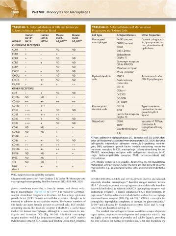
                TABLE 68–1.  Selected Markers of Different Monocyte    TABLE 68–2.  Selected Markers of Mononuclear
                Subsets in Mouse and Human Blood                       Phagocytes and Related Cells
                         Human      Human     Mouse      Mouse         Cell Type        Antigen Markers  Other Properties
                             hi
                         CD14       CD14+     CCR2+      CCR2–         Monocytes/       F4/80 (mouse)    Opsonic phagocyto-
                Antigen  CD16–      CD16+     CX CR1 low  CX CR1 hi
                                                3          3           macrophages      EMR2 (human)     sis; lysozyme secre-
                CHEMOKINE RECEPTORS                                                     CD68             tion; abundant acid
                CCR1     +          –         ND         ND                             CR3 (CD11b)      hydrolases
                CCR2     +          –         +          –                              Sialoadhesin        
                CCR4     +          –         ND         ND                             (Siglec-1)        
                CCR5     –          +         ND         ND                             Scavenger receptors
                                                                                        (SR-A, MARCO)
                CCR7     +          –         ND         ND                             Mannose receptor
                CXCR1    +          –         ND         ND                             M-CSF receptor
                CXCR2    +          –         ND         ND            Myeloid dendritic   MHC II        Activation of naïve
                CXCR4    +          ++        ND         ND            cells            Costimulatory    CD4 T lymphocytes
                CX CR1   +          ++        +          ++                             molecules
                  3                                                                     CD11c
                OTHER RECEPTORS
                                                                                        CD8α+/–
                CD4      +          +         ND         ND
                                                                                        DEC205
                CD11a    ND         ND        +          ++                             DC-SIGN
                CD11b    ++         ++        ++         ++                             DC-LAMP
                CD11c    ++         +++       –          +             Plasmacytoid     CD123            Type I interferon
                CD14     +++        +         ND         ND            dendritic cells  B220             production; in vitro
                                                                                                         growth by flt-3
                CD31     +++        +++       ++         +                              Lectin-like receptors   ligand
                                                                                        (Siglec-H)
                CD32     +++        +         ND         ND
                                                                                                                 +
                                                                       Osteoclasts      CD68             Vacuolar H  ATPase;
                CD33     +++        +         ND         ND
                                                                                        TRAP             proteinase K;
                CD43     ND         ND        –          +                                               resorption of living
                                                                                        Calcitonin receptor  bone
                CD49b    ND         ND        +          –                              α β
                CD62L    ++         –         +          –                               v 3
                                                                      ATPase, adenosine triphosphatase; DC, dendritic cell; DC-LAMP, den-
                CD86     +          ++        ND         ND           dritic cell lysosomal-associated membrane protein; DC-SIGN, dendritic
                                                                      cell–specific intercellular adhesion molecule-3–grabbing noninte-
                CD115    ++         ++        ++         ++
                                                                      grin; EMR, epidermal growth factor module-containing mucin-like
                CD116    ++         ++        ++         ++           hormone receptor; M-CSF, macrophage colony-stimulating  factor;
                F4/80    ND         ND        +          +            MARCO, macrophage receptor with collagenous structure; MHC,
                                                                      major  histocompatibility  complex;  TRAP,  tartrate-resistant acid
                Ly6C     ND         ND        +          –            phosphatase.
                7/4      ND         ND        +          –            note: Marker expression is variable, depending on cell localization,
                                                                      maturation, and activation. Some markers are also present on other
                MHC      +          ++        –          –            myeloid cells, e.g., polymorphonuclear cells, and selected endothelial
                class II
                                                                      cells.
               MHC, major histocompatibility complex.
               Adapted with permssion from Gordon S. & Taylor PR: Monocyte and   CD11b/CD18 (Mac1, CR3), and CD11c, present on DCs and selected,
               macrophage heterogeneity. Nat Rev Immunol 5(12):953–964, 2005.  especially alveolar, macrophages.  Receptor antigen markers include
                                                                                              50
                                                                      SR-A,  a broadly expressed macrophage receptor additionally found on
                                                                          51
               plasma membrane molecules, is broadly present and almost exclu-  sinusoidal endothelium, whereas MARCO (macrophage receptor with
               sive to macrophages (Fig. 68–5A to C). 48,49  It is related to G-protein–   collagenous structure), a related collagenous SR, is more restricted in
                                                                              52
               coupled chemokine receptors in structure, but has a large epidermal   expression.  Additional markers include lectins such as the macrophage
                                                                                                       53
               growth factor (EGF) domain extracellular extension, thought to be   mannose/fucose receptor (MR; Fig. 68–5E).  CD163, a receptor for
                                                                                                                        54
               involved in adhesion to extracellular matrix. The human members of   hemoglobin–haptoglobin complexes, is induced by glucocorticoids,
                                                                                        55
               this family are more broadly present on myeloid cells; EGF module-  IL-10,  and substance P.  Complement receptors (CRs) and Fc recep-
                                                                          54
               containing mucin-like hormone receptor 2 (EMR2) is a useful tissue   tors (FcRs) are described in Chap. 67.
               marker for human macrophages, although it is also present in neu-  The resident macrophages in tissues constitute a major dispersed
               trophils and immature DCs (Fig. 68–5A). Additional macrophage   organ system, responsive to endogenous and exogenous stimuli; they
               antigen markers useful for immunocytochemical and FACS analysis   are highly active in uptake of particles and soluble ligands, providing
               include Siglec1 (Fig. 68–5D), a sialic acid-binding lectin, the β  integrins   not only sentinels for defense at portals of entry, but also mediating the
                                                           2


          Kaushansky_chapter 68_p1075-1088.indd   1080                                                                  9/17/15   3:41 PM
   1100   1101   1102   1103   1104   1105   1106   1107   1108   1109   1110