
Page 1110 - Williams Hematology ( PDFDrive )
P. 1110
1084 Part VIII: Monocytes and Macrophages Chapter 68: Production, Distribution, and Activation of Monocytes and Macrophages 1085
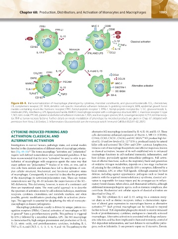
Figure 68–9. Immunomodulation of macrophage phenotype by cytokines, microbial constituents, and glucocorticosteroids. CCL, chemokines;
CR, complement receptor; DC-SIGN, dendritic cell–specific intercellular adhesion molecule-3–grabbing nonintegrin; EMR, epidermal growth factor
module-containing mucin-like hormone receptor; FPR1, formyl-peptide receptor 1; FPRL-1, formyl-peptide receptor-like 1; GC, glucocorticoids; IL,
interleukin; INFγ, interferon-γ; LPS, lipopolysaccharide; MARCO, macrophage receptor with a collagenous structure; MRC-1, mannose receptor C-type
1; NO, nitric oxide; PECAM, platelet endothelial cell adhesion molecule-1; ROS, reactive oxygen species; SR-A, scavenger receptor A; TLR, toll-like recep-
tor; TNF-α, tumor necrosis factor-α. Further details on innate modulation of phenotype by microbial products are given in Chap. 67. (Adapted with
permission from Yona, S. & Gordon, S.: Inflammation: Glucocorticoids turn the monocyte switch. Immunol Cell Biol 85(2):81–82, 2007.)
CYTOKINE-INDUCED PRIMING AND alternative M2 macrophage is mediated by IL-4, IL-10, and IL-13. These
ACTIVATION: CLASSICAL AND cells demonstrate enhanced expression of Dectin-1, MR C1 (CD206),
CD163, CCR2, CXCR1, CXCR2, and DC-SIGN. M2 produce high lev-
84
ALTERNATIVE ACTIVATION els of IL-10 and low-levels of IL-12. IFN-γ, produced mainly by natural
85
Investigations in normal humans, pathologic states, and animal models killer cells and activated Th1 CD4+ and CD8+ cytotoxic lymphocytes,
have led to the characterization of different states of macrophage polariza- induces a set of macrophage biosynthetic and effector responses, known
tion (Fig. 68–10). The terms macrophage “activation and “polarization” as classical activation, because of its well-established role in enhanced
81
require well-defined nomenclature and experimental guidelines. It has macrophage functions in cell-mediated immunity, inflammation, and
been recommended that the term “activation” be used to refer to per- host defense, particularly against intracellular pathogens. Full activa-
turbation of macrophages with exogenous agents the same way that tion of effector functions, such as the respiratory burst and generation
many authors use “polarization.” Studies of in vitro, ex vivo, and in of oxidative nitrogen metabolites, depends on a two-stage mechanism
82
vivo cells from animals and humans have led to descriptions of com- of priming by the cytokine, via specific IFN-γ receptors, followed by a
plex cellular structural, biochemical, and functional activation states local stimulus, LPS, or other TLR ligands. Although essential for host
of macrophages. Consequently, it is essential to describe the properties defense, including against opportunistic pathogens such as found in
of the macrophage its activation/polarization state. There are various patients with the acquired immunodeficiency syndrome, classical acti-
nomenclatures and Fig. 68–10 indicates the major features of the M1– vation is responsible for tissue injury and its consequences in inflam-
M2 dichotomy. This dichotomy, however, is more of a continuum and matory bowel disease, tuberculosis, and rheumatoid arthritis, although
there are transitional states. The most useful approach is to describe additional immunopathogenic agents, such as immune complexes, also
the spectrum of activation states by cell isolation technique, membrane contribute. Biochemical and cellular aspects of classical activation are
receptors, cytokines, chemokines, and metabolic markers, and when described in Chap. 67.
possible, genetic modifications producing shifts in activation pheno- The Th2 cytokines IL-4 and IL-13, acting via a common recep-
type. This approach is essential for deciphering the role of monocyte– tor chain as well as distinct receptors, induce a characteristic signa-
macrophages in disease pathogenesis. ture of altered gene expression in macrophages known as alternative
Macrophage polarization usually is driven by unique pattern rec- activation. 86,87 Such primed macrophages can be induced to respond
ognition receptors (PRRs). Classical activation of macrophages or M1 further to local, TLR-dependent phagocytic stimuli to secrete enhanced
in general have a proinflammatory profile. This pathway is triggered levels of proinflammatory cytokines, analogous to classically activated
83
by IFN-γ followed by a microbial stimulus, LPS. The M1 macrophage macrophages. Alternative activation is associated with allergy and para-
is characterized by high antigen presentation and production of IL-12, sitic infection, and has been implicated in humoral immunity, control of
IL-23, nitric oxide, and proinflammatory cytokines, including IL-1, Th1-dependent inflammation, and host defense to extracellular patho-
TNF-α, IL-6, and CXCL-1, -2, -3, -5, -8, -9, and -10. The pathway to the gens, such as helminths. It can promote repair or, if excessive, fibrosis.
Kaushansky_chapter 68_p1075-1088.indd 1085 9/17/15 3:42 PM