
Page 1337 - Williams Hematology ( PDFDrive )
P. 1337
1312 Part X: Malignant Myeloid Diseases Chapter 85: Essential Thrombocythemia 1313
number, the megakaryocytes are generally small and hypolobulated, in
60
TABLE 85–2. Causes of Thrombocytosis contrast to the large hyperlobulated forms typical of ET. A raised plate-
CLONAL THROMBOCYTOSIS let count is also a feature of refractory anemia with ringed sideroblasts
and thrombocytosis (RARS-T), and may be associated with thrombotic
Essential thrombocythemia complications. Approximately half of patients with RARS-T harbor a
Polycythemia vera JAK2 V617F mutation or, rarely, a mutation in MPL.
Primary myelofibrosis
Chronic myeloid leukemia PATHOGENETIC RELATIONSHIP OF
Refractory anemia with ringed sideroblasts and thrombocytosis ESSENTIAL THROMBOCYTHEMIA TO OTHER
5q-minus syndrome MYELOPROLIFERATIVE NEOPLASMS
REACTIVE (SECONDARY) THROMBOCYTOSIS
Polycythemia Vera
Transient thrombocytosis The same JAK2 V617F mutation is present in the vast majority of patients
Acute blood loss with PV (Chap. 84) and in approximately half of those with ET, rais-
Recovery from thrombocytopenia (rebound thrombocytosis) ing questions as to how a single mutation is commonly associated with
Acute infection or inflammation apparently distinct clinical phenotypes. Clones that are homozygous
Response to exercise for the JAK2 V617F mutation (arising by a mitotic recombination event
Response to drugs (vincristine, epinephrine, all-trans-retinoic acid) termed uniparental disomy; Fig. 85–5) are larger and more frequent in
suggesting a role for increased
patients with PV compared with ET,
62,63
Sustained thrombocytosis JAK2-STAT5 signaling in driving erythrocytosis. In support of this
Iron deficiency hypothesis, in both mouse and human model systems strong JAK2-
Splenectomy or congenital absence of spleen STAT5 activation drives erythropoiesis whereas weaker activation
Malignancy favors a megakaryopoiesis. 16,64,65 Other contributing factors include the
63
Chronic infection or inflammation effects of patient gender and modulation of STAT1 signaling. 66
Hemolytic anemia Myelofibrosis and Accelerated Phase Disease
FAMILIAL THROMBOCYTOSIS A proportion of patients diagnosed with ET experience progres-
SPURIOUS THROMBOCYTOSIS sion to an accelerated phase characterized by increasingly disordered
hematopoiesis. The phenotypic manifestations are variable and include
Cryoglobulinemia hyperproliferation, myelodysplasia, or, most commonly, myelofibrosis.
Cytoplasmic fragmentation in acute leukemia Myelofibrotic transformation of ET, characterized by marrow fibrosis,
Red cell fragmentation extramedullary hematopoiesis and marrow failure, is clinically indis-
Bacteremia tinguishable from PMF (Chap. 86), suggesting PMF may represent pre-
sentation with accelerated phase disease. Consistent with this, patients
with PMF may have thrombocytosis for many years prior to diagnosis,
in ET. Given the significant impact of tyrosine kinase inhibitors on the suggestive of undiagnosed ET. 58
prognosis of CML, it is important that this unusual presentation is not The prevalence of mutations in JAK2, CALR, or MPL is similar in
overlooked. It is therefore recommended that suspected cases of ET that ET compared to myelofibrosis; however, karyotypic abnormalities are
are negative for a relevant somatic mutation undergo molecular analysis present in up to 50 percent of myelofibrosis patients (Chap. 86) com-
of blood for the BCR-ABL1 fusion gene. Marrow aspiration, biopsy and pared to only approximately 5 percent of patients in ET, indicating a
G-banding cytogenetic analysis, may be useful in a specific case. greater degree of genetic instability. In addition, mutations in genes
implicated in transcriptional regulation (including ASXL1, IDH1/2,
and EZH2) appear more common in patients with PMF compared
MYELODYSPLASIA with ET. Together, these findings suggest that progression to advanced
Thrombocytosis, usually in association with anemia, may be seen in phase disease arises through a process of clonal evolution driven by the
the myelodysplastic disorder associated with an isolated deletion of acquisition of additional genetic events or epigenetic alterations; to date,
chromosome 5q (“5q-minus syndrome”). Although often increased in however, no combination of genetic events has been shown to reliably
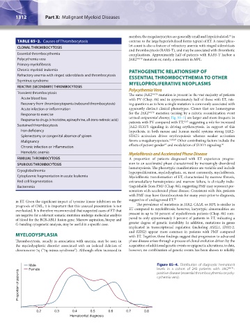
Male Figure 85–4. Distribution of diagnostic hematocrit
Female levels in a cohort of 243 patients with JAK2 V617F -
Proportion of patients
positive disease (essential thrombocythemia or poly-
cythemia vera).
0.2 0.3 0.4 0.5 0.6 0.7 0.8
Hematocritat diagnosis
Kaushansky_chapter 85_p1307-1318.indd 1312 9/21/15 11:08 AM