
Page 1602 - Williams Hematology ( PDFDrive )
P. 1602
1576 Part XI: Malignant Lymphoid Diseases Chapter 95: General Considerations for Lymphomas 1577
This translocation has been identified in approximately 50 percent
of systemic cases in adults and in a majority of pediatric cases of
ALCL. 142,143 The t(2;5) translocation is rare in primary cutaneous ALCL,
which is generally considered to be a different disease than systemic
ALCL (Chap2. 103 and 104.) 144
MARGINAL ZONE LYMPHOMA OF MUCOSA-
ASSOCIATED LYMPHATIC TISSUE
t(11;18)(API2-MALT1), t(1;14)(IGH-BCL10), t(14;18)(IGH-MALT1),
and t(3;14)(IGH-FOXP1) occur in marginal zone B-cell lymphoma of
MALT of different sites. The first three chromosome translocations are
specifically associated with the marginal zone lymphoma of MALT lym-
phomas and the oncogenic products of these translocations target the
nuclear factor-κB pathway (Chap. 101). 101
MANTLE CELL LYMPHOMA
t(11;14)(q13;q32) is present in the cells of most cases of mantle cell lym-
phoma and results in cyclin D1 upregulation. iFISH is the most useful
test to identify the juxtaposition of the CCND1 and IGH genes in mantle
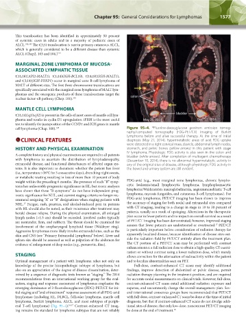
cell lymphoma (Chap. 100). 145 Figure 95–4. Fluorine-deoxyglucose positron emission tomog-
18
raphy/computed tomography (FDG-PET/CT) imaging of Burkitt
lymphoma before and after successful therapy. At the time of initial
CLINICAL FEATURES diagnosis (May 21, 2014), hypermetabolic areas of avid FDG uptake
were detected in a right cervical mass, clavicle, abdominal lymph nodes,
HISTORY AND PHYSICAL EXAMINATION stomach, and pelvic bones (yellow arrows) in this patient with stage
A complete history and physical examination are required in all patients IV lymphoma. Physiologic FDG activity is also seen in the colon and
bladder (white arrows). After completion of multiagent chemotherapy
with lymphoma to ascertain the distribution of lymphadenopathy, (December 10, 2014), there is no abnormal hypermetabolic activity in
extranodal disease, and functional disturbances of affected organ sys- any of the original sites of disease, although physiologic FDG activity in
tems. It is also important to document whether the patient has fever the bowel and urinary system are still evident.
(i.e., temperature >38°C for 3 consecutive days), drenching night sweats,
or metabolic wasting resulting in loss of more than 10 percent of body
weight within the preceding 6 months. The presence of such “B” symp- FDG-avid (e.g., most marginal zone lymphomas, chronic lympho-
toms has unfavorable prognostic significance in HL, but recent analyses cytic leukemia/small lymphocytic lymphoma, lymphoplasmacytic
have shown that these “B symptoms” do not have independent prog- lymphoma/ Waldenström macroglobulinemia, angioimmunoblastic T-cell
nostic significance for NHL, and current staging criteria no longer rec- lymphoma, mycosis fungoides, and cutaneous B-cell lymphomas). For
ommend assigning “A” or “B” designations when staging patients with FDG-avid lymphomas, PET/CT imaging has been shown to improve
17
NHL. Fatigue, rash, pruritus, and alcohol-induced pain in patients the accuracy of staging for both nodal and extranodal sites compared
with HL should also be noted, as their recurrence after treatment may with CT imaging, leading to a change in stage in 10 to 30 percent of
herald disease relapse. During the physical examination, all enlarged patients, usually as a result of upstaging. Alterations in the therapeutic
lymph nodes (>1.5 cm) should be recorded. Involved nodes typically plan occur in fewer patients and no impact on overall survival as a result
are nontender, firm, and rubbery. The throat should be examined for of PET/CT imaging has been demonstrated; however, improved staging
17
involvement of the oropharyngeal lymphoid tissue (Waldeyer ring). assures that fewer patients are undertreated or overtreated. PET/CT
Aggressive lymphomas more likely involve extranodal sites, such as the is particularly important before consideration of radiation therapy for
skin and CNS (see “Primary Extranodal Lymphoma” below). Liver and apparently localized disease, because identification of disease sites out-
spleen size should be assessed as well as palpation of the abdomen for side the radiation field by PET/CT entirely alters the treatment plan.
evidence of enlargement of deep nodes (e.g., paraaortic, iliac). The CT portion of a PET/CT scan may be performed with contrast
enhancement at a full radiation dose to obtain a high-quality CT exami-
nation or without contrast using a lower radiation dose, which merely
STAGING allows correction for the attenuation of radioactivity within the patient
Optimal management of a patient with lymphoma relies not only on and to localize abnormalities seen on PET.
knowledge of the precise histopathologic subtype of lymphoma, but Full-dose, contrast-enhanced CT scans may identify additional
also on an appreciation of the degree of disease dissemination, deter- findings, improve detection of abdominal or pelvic disease, permit
mined by a sequence of diagnostic tests known as “staging.” The 2014 radiation therapy planning in the treatment position, and are required
recommendations from an international working group for the eval- for accurate nodal measurements on clinical trials. However, full-dose,
uation, staging and response assessment of lymphomas emphasize the contrast-enhanced CT scans entail additional radiation exposure and
emerging dominance of 2-fluorodeoxyglucose (FDG)-PET/CT for ini- expense, and uncommonly change the overall management plan. Sev-
tial staging and “end of treatment” response assessment of all FDG-avid eral international consensus groups have recommended that PET/CT
lymphomas (including HL, DLBCL, follicular lymphoma, mantle cell with full-dose, contrast-enhanced CT scans be done at the time of initial
lymphoma, Burkitt lymphoma, ALCL, and most subtypes of periph- diagnosis, but that if contrast-enhanced CT scans do not divulge addi-
eral T-cell lymphoma) (Fig. 95–4). 16,17 Contrast-enhanced CT imag- tional sites of disease, that only low-dose, noncontrast PET/CT imaging
ing remains the standard for lymphoma subtypes that are not reliably be done at the end of treatment. 16
Kaushansky_chapter 95_p1569-1586.indd 1577 9/21/15 12:17 PM