
Page 1998 - Williams Hematology ( PDFDrive )
P. 1998
1972 Part XII: Hemostasis and Thrombosis Chapter 115: Vascular Function In Hemostasis 1973
muscles cells, and macrophages, which express plasminogen activators, In vivo, the circulating half-life of t-PA is approximately 5 min-
plasminogen activator inhibitors, and fibrinolytic receptors. utes. Infusion of DDAVP, bradykinin, platelet-activating factor (PAF),
endothelin, or thrombin is associated with an acute release of t-PA,
123
ENDOTHELIAL CELL PRODUCTION OF and a burst of fibrinolytic activity can be detected within minutes. In
FIBRINOLYTIC PROTEINS the mouse lung, exposure to hyperoxia leads to 4.5-fold upregulation
107
of t-PA mRNA in small-vessel endothelial cells. In humans, infusion
In 1958, Todd demonstrated that human blood vessels possess fibrino- of TNF into patients with malignancy is associated with an increase in
lytic activity that is dependent upon an intact endothelium. 102,103 We plasma t-PA. Deficient release of t-PA in response to venous occlusion
123
124
now know that the endothelium is the principal source of t-PA in vivo in humans is associated with deep venous thrombosis, as well as atro-
where it appears to be highly restricted to small blood vessels in specific phie blanche and other cutaneous vasculitides. 125
anatomic locations, a pattern that likely reflects the heterogeneity of In vivo, urokinase plasminogen activator (u-PA) is not a product
126
endothelial cells as they respond to a myriad of tissue-specific cues. 104,105 of resting endothelium, but is produced primarily by renal tubular
127
In the baboon, for example, sites of t-PA production include 7 to 30 μm epithelium. Expression of u-PA mRNA in endothelium, however, is
precapillary arterioles and postcapillary venules, but not large arteries strongly stimulated during wound repair and physiologic angiogenesis
106
and veins. In the mouse lung, similarly, bronchial, but not pulmonary, within ovarian follicles, corpus luteum, and maternal decidua. Endo-
128
107
129
endothelial cells express t-PA. Moreover, enhanced expression of t-PA thelial cells passaged in culture do synthesize u-PA, and expression of
130
at branch points of pulmonary blood vessels may reflect stimulation by its mRNA is stimulated by TNF-α by 5- to 30-fold. Small increases in
laminar shear stress. In addition, peripheral sympathetic neurons that u-PA have also been observed in vitro in response to IL-1 and LPS. 131–133
108
invest the walls of small arteries may represent a significant source of The association of u-PA with the blood vessel wall appears to reflect
circulating t-PA. 109 its association with the u-PA receptor (uPAR) which may fulfill a variety
Although in vitro studies suggest that t-PA expression in cultured of nonproteolytic functions ranging from directed cell migration to cel-
endothelial cells is regulated by a wide array of factors, only a few of these lular adhesion, differentiation, and proliferation (Fig. 115–6). In the
134
pathways have been confirmed in vivo. Thrombin, histamine, 111,112 adult mouse, uPAR mRNA is not normally detected by in situ hybrid-
110
113
135
oxygen radicals, phorbol myristate acetate, DDAVP (deamino ization in the endothelium of either large or small blood vessels.
114
115
D-arginine vasopressin), and butyric acid liberated from dibutyryl However, upon stimulation with endotoxin, expression is detected in
cAMP all increase t-PA mRNA in cultured endothelial cells. Both endothelium lining aorta, as well as arteries, veins, and capillaries in
116
135
thrombin and histamine appear to act via receptor-mediated activation heart, kidney, brain, and liver, and in renal tubular epithelial cells. 127
105
of the protein kinase C pathway. Laminar shear stress stimulates both Plasminogen activator inhibitor (PAI)-1 is likely to function as a
118
t-PA secretion and steady-state mRNA levels. Hyperosmotic stress major regulator of plasmin generation in the vicinity of the endothelial
117
and repetitive stretch also enhance t-PA expression. 119,120 In addition, cell. Thrombin, IL-1, transforming growth factor β, TNF, lipoprotein(a)
differentiating agents, such as retinoids, 121,122 stimulate transcription of (Lp[a]), and LPS all induce dramatic increases in steady state PAI-1
t-PA in endothelial cells in vitro. message levels. 110,131,132,136,137 Heparin-binding growth factor 1 reduces
Plasmin
t-PA t-PA
A-tail A-tail
PLG PLG
u-PA
A 2
A 1 A 2 A 3 A 4 A 1 A 2 A 3 A 4 U 1
p11 p11 uPAR
U
U 3 2
A Plasma membrane B Plasma membrane
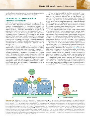
Figure 115–6. Schematic of principal endothelial cell fibrinolytic receptors. A. The annexin A2/S100A10 heterotetrameric complex. Annexin A2
consists of a hydrophilic aminoterminal tail domain (A-Tail, approximately 3 kDa), and a membrane-oriented carboxyl terminal core domain (approxi-
mately 33 kDa). 311,312 The tail domain contains residues required for tissue-type plasminogen activator (t-PA) binding. The core domain is composed of
four homologous annexin repeats (A1, A2, A3, and A4), each consisting of five α-helical regions that contribute to calcium-dependent phospholipid
binding sites. Repeat 2 appears to be most important for the interaction of annexin A2 with the endothelial cell surface. Plasminogen (PLG) binding
requires lysine residue 307 within helix C of repeat 4. B. Urokinase plasminogen activator receptor (uPAR) is a 55- to 60-kDa, glycosylphosphatidyli-
nositol-linked protein that consists of three disulfide-linked domains (U1, U2, U3). Domain 1 contains sequences required for urokinase plasmino-
314
gen activator (u-PA) binding, while domains 2 and 3 mediate the receptor’s interaction with matrix proteins such as vitronectin. Domain 3 contains
glycosylphosphatidylinositol-linked membrane anchor. (A, adapted with permission from Gerke V, Creutz CE, Moss SE: Annexins: linking Ca2+ signalling to
membrane dynamics. Nat Rev Mol Cell Biol 6(6):449–461.)
Kaushansky_chapter 115_p1967-1984.indd 1973 9/18/15 10:08 AM