
Page 439 - Williams Hematology ( PDFDrive )
P. 439
414 Part V: Therapeutic Principles Chapter 26: Immune Cell Therapy 415
engraftment of human leukemia in nonobese diabetic/severe combined CTL selectively lysed leukemic blasts and prevented engraftment of
immunodeficiency mice, providing evidence that the leukemic stem cell leukemia in immunodeficient mice, suggesting that these T cells may
can be recognized by allogeneic T cells. 126 mediate an antileukemic effect without affecting normal hematopoie-
Most mHAgs result from nonsynonymous single nucleotide poly- sis in vivo. 140,144 Recent work has identified WT-1–specific T cells after
morphisms (SNPs) in the coding sequence of donor and recipient genes HLA-identical sibling HSCT and correlated these cells with a GVL
145
that alter the HLA binding or TCR contact of HLA-bound peptides. effect. In a recent pilot trial, 11 high-risk leukemia patients received
146
There are several million SNPs with an allele frequency of greater than infusions of WT-1–specific T cells. In four of these patients, the
5 percent in the human genome, including approximately 50,000 SNPs infused T cells were generated in the presence of IL-21, which mod-
127
that lead to amino acid changes in proteins. Identification of the poly- ulates the differentiation of T cells in culture. The transferred T cells
morphic genes that encode mHAgs is facilitated by the data on genetic exhibited evidence of leukemic activity, and CTL that were generated
variation from the human HapMap project, which has enabled the use with IL-21 had superior antileukemic activity and survived long-term
of whole-genome association analysis for mHAg discovery in addition as memory T cells. Additional study in a larger number of patients is
146
123
to conventional techniques for antigen discovery. Identifying the needed, but these results offer a potential safe and effective treatment for
subset of mHAgs that will be the most useful to target to augment the patients with limited treatment options.
GVL effect requires consideration of several factors, including the allele
frequency of the mHAg encoding gene, the HLA restricting allele that GENETIC RETARGETING OF T CELLS
presents the mHAg, and the expression and presentation of the mHAg
128
in leukemia cells and nonhematopoietic tissues. Most mHAg discov- Extending cellular therapy to patients from whom tumor-reactive T
ery efforts have focused on CD8+ T cells, but CD4+ T cells are likely cells cannot be isolated and to other malignancies can be accomplished
to play a key role either as direct effector cells in the GVL response, or to by using gene transfer approaches to retarget patient T cells to recog-
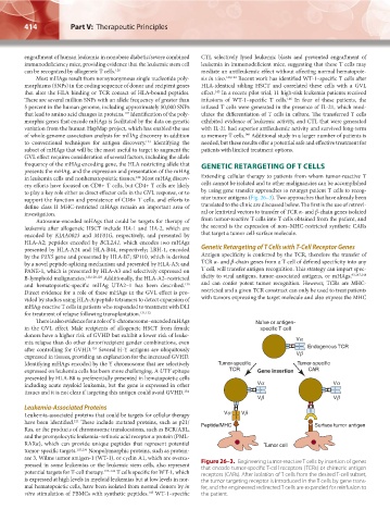
support the function and persistence of CD8+ T cells, and efforts to nize tumor antigens (Fig. 26–3). Two approaches that have already been
define class II MHC-restricted mHAgs remain an important area of translated to the clinic are discussed below. The first is the use of retrovi-
investigation. ral or lentiviral vectors to transfer of TCR α- and β-chain genes isolated
Autosome-encoded mHAgs that could be targets for therapy of from tumor-reactive T cells into T cells obtained from the patient, and
leukemia after allogeneic HSCT include HA-1 and HA-2, which are the second is the expression of non–MHC-restricted synthetic CARs
encoded by KIAA0023 and MY01G, respectively, and presented by that target a tumor cell-surface molecule.
HLA-A2; peptides encoded by BCL2A1, which encodes two mHAgs
presented by HLA-A24 and HLA-B44, respectively; LRH-1, encoded Genetic Retargeting of T Cells with T-Cell Receptor Genes
by the P2X5 gene and presented by HLA-B7; SP110, which is derived Antigen specificity is conferred by the TCR, therefore the transfer of
by a novel peptide-splicing mechanism and presented by HLA-A3; and TCR α- and β-chain genes from a T cell of defined specificity into any
PANE-1, which is presented by HLA-A3 and selectively expressed on T cell, will transfer antigen recognition. This strategy can impart spec-
B-lymphoid malignancies. 123,128,129 Additionally, the HLA-A2–restricted ificity to viral antigens, tumor-associated antigens, or mHAgs, 93,147,148
and hematopoietic-specific mHAg UTA2–1 has been described. and can confer potent tumor recognition. However, TCRs are MHC-
130
Direct evidence for a role of these mHAgs in the GVL effect is pro- restricted and a given TCR construct can only be used to treat patients
vided by studies using HLA-A/peptide tetramers to detect expansion of with tumors expressing the target molecule and also express the MHC
mHAg-reactive T cells in patients who responded to treatment with DLI
for treatment of relapse following transplantation. 131,132
There is also evidence for a role of Y-chromosome–encoded mHAgs Naïve or antigen-
in the GVL effect. Male recipients of allogeneic HSCT from female specific T cell
donors have a higher risk of GVHD but exhibit a lower risk of leuke-
mia relapse than do other donor/recipient gender combinations, even Va
133
after controlling for GVHD. Several H-Y antigens are ubiquitously Endogenous TCR
expressed in tissues, providing an explanation for the increased GVHD. Vb
Identifying mHAgs encoded by the Y chromosome that are selectively Tumor-specific Tumor-specific
expressed on leukemia cells has been more challenging. A UTY epitope TCR Gene insertion CAR
presented by HLA-B8 is preferentially presented in hematopoietic cells
including acute myeloid leukemia, but the gene is expressed in other Va Va
tissues and it is not clear if targeting this antigen could avoid GVHD. 134
Vb Vb
Leukemia-Associated Proteins
Leukemia-associated proteins that could be targets for cellular therapy Va Vb
121
have been identified. These include mutated proteins, such as p21/ Peptide/MHC Surface tumor antigen
Ras, or the products of chromosome translocations, such as BCR/ABL,
and the promyelocytic leukemia–retinoic acid receptor α protein (PML-
RARα), which can provide unique peptides that represent potential Tumor cell
tumor-specific targets. 135,136 Nonpolymorphic proteins, such as protein-
ase 3, Wilms tumor antigen-1 (WT-1), or cyclin A1, which are overex-
pressed in some leukemias or the leukemic stem cells, also represent Figure 26–3. Engineering tumor-reactive T cells by insertion of genes
that encode tumor-specific T-cell receptors (TCRs) or chimeric antigen
potential targets for T-cell therapy. 137–142 T cells specific for WT-1, which receptors (CARs). After isolation of T cells from the desired T-cell subset,
is expressed at high levels in myeloid leukemias but at low levels in nor- the tumor targeting receptor is introduced in the T cells by gene trans-
mal hematopoietic cells, have been isolated from normal donors by in fer, and the engineered redirected T cells are expanded for reinfusion to
143
vitro stimulation of PBMCs with synthetic peptides. WT-1–specific the patient.
Kaushansky_chapter 26_p0409-0420.indd 414 9/17/15 6:01 PM