
Page 690 - Williams Hematology ( PDFDrive )
P. 690
664 Part VI: The Erythrocyte Chapter 46: Erythrocyte Membrane Disorders 665
3 as part of a macromolecular complex, and may serve as a chaperone Linker proteins mediate the vertical attachment of the skeleton to inte-
for band 3 targeting to the membrane. GPC associates with protein gral membrane proteins in the lipid bilayer (see Fig. 46–1). The primary
25
4.1R and p55, thereby providing an additional contact site between the connecting protein is ankyrin, which links spectrin to the cytoplasmic
membrane and the skeleton (see Fig. 46–1). These interactions play a domain of band 3, as well as to the Rh–RhAG complex. Protein 4.1R
19
role in stabilizing the membrane. provides an additional link with GPC and band 3.
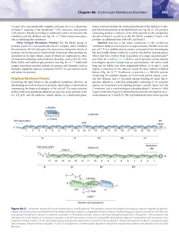
Other Integral Membrane Proteins The Rh-RhAG group of Spectrin Spectrin is the major constituent of the erythrocyte
proteins is part of a macromolecular band 3 complex, which stabilizes membrane skeleton and is present at approximately 240,000 molecules
the membrane. RhAG belongs to the ammonium transporter family of per cell. It is a multifunctional protein composed of two homologous
29
proteins, but its function is controversial. Numerous other proteins are but structurally distinct subunits, α and β, encoded by separate genes,
embedded in the lipid bilayer, many of which are implicated in clini- which may have evolved from duplication of a single ancestral gene
30
cal immunohematology and membrane disorders, such as the XK, Kell, (see Table 46–1 and Fig. 46–3). Both α- and β-spectrin contain tandem
Kidd, Duffy, and Lutheran glycoproteins (see Fig. 46–1). 11,19 Additional homologous spectrin repeats that are approximately 106 amino acids
integral membrane proteins include ion pumps and channels, such as long and are folded into three antiparallel helices, A, B, and C. Each
stomatin, aquaporin, glucose transporter (GLUT-1), and various cation repeat is connected to the adjacent repeat by short ordered α-helical
and anion transporters. linkers (Fig. 46–3). 31,32 Erythrocyte α-spectrin is a 280-kDa protein
comprising 20 complete repeats, an N-terminal partial repeat, a cen-
Peripheral Membrane Proteins tral SH3 domain, and a C-terminal calcium-binding EF hand. The β-
Underlying the lipid bilayer is the peripheral membrane skeleton, an spectrin subunit is a 246-kDa polypeptide consisting of 16 complete
interlocking network of structural proteins, which plays a critical role in repeats, an N-terminal actin binding domain, a partial repeat near the
maintaining the shape and integrity of the red cell. The major proteins C-terminus, and a nonhomologous phosphorylated C-terminus. Mild
of the erythrocyte membrane skeleton are spectrin, actin, proteins 4.1R, trypsin treatment of spectrin cleaves the two subunits into distinct struc-
4.2, 4.9, p55, and the adducins, which interact in a horizontal plane. tural domains: αI-V and βI-IV. The triple helical structure of the spectrin
α B-Helix
α A-Helix a-Spectrin
α C-Helix
Self-association EF
SH3 hands
N C
1 2 3 4 5 6 7 8 9 10 11 12 13 14 15 16 17 18 19 20 21
αl αll αlll αlV αV
Self-association b-Spectrin Nucleation
Ankyrin site
C P P
P
P 4.1 N
17 16 15 14 13 12 11 10 9 8 7 6 5 4 3 2 1
βl βll βlll βlV
C P P
P β B-Helix
P
β
β A-Helix α
β16 α1
N α C-Helix
α0/β17
Spectrin self-association
Figure 46–3. Schematic model of human erythrocyte α- and β-spectrin. The proteins consist of multiple homologous spectrin repeats of approxi-
mately 106 amino acids numbered from the N-terminal. Each repeat is composed of three α helices. Nonhomologous regions include an SH3 domain
and calcium-binding EF hands in α-spectrin, a protein 4.1R binding domain, and a C-terminal phosphorylated tail in β-spectrin. The nucleation site
indicates the initial region of interaction between α and β monomers to form an antiparallel heterodimer. Spectrin heterodimer self association into
tetramers involves helix C of the α0 partial repeat of α-spectrin and helices A and B of the partial β17 repeat of β-spectrin to form a complete triple
helical repeat. Ankyrin binds to repeats 14 and 15 of β-spectrin. Limited tryptic digestion of spectrin cleaves the proteins into discrete αI-V and βI-IV
domains.
Kaushansky_chapter 46_p0661-0688.indd 665 9/17/15 6:41 PM