
Page 1011 - Clinical Immunology_ Principles and Practice ( PDFDrive )
P. 1011
CHaPter 72 Immunological Lung Diseases 975
disorders, including CTDs, malignancy, infections, and those opacities can be migratory and usually have a peripheral distribu-
caused by medications. tion similar to those seen in chronic eosinophilic pneumonia.
Rarer radiographic manifestations include linear or nodular
Clinical Manifestations interstitial opacities and honeycombing. The presence of a pleural
The onset of disease is usually in the fifth to sixth decades of effusion or pleural thickening should suggest an associated CTD.
life; men and women are equally affected. Most individuals have HRCT shows patchy airspace consolidation, especially in the
symptoms for less than 2 months before diagnosis. The initial lung periphery with a lower-lung zone predominance (see Fig.
presentation is usually with a dry, nonproductive cough and 72.10B). Other findings include ground-glass attenuation, small
flu-like symptoms, including fever, sore throat, and malaise. This nodular opacities, and bronchial wall thickening.
is followed by progressive dyspnea, and routine laboratory As in other ILDs, a restrictive ventilatory defect is the most
evaluation is nonspecific. common pulmonary function abnormality. Gas exchange
Chest radiography shows diffuse, often patchy alveolar opacities abnormalities are common and are accompanied by decreased
in the setting of normal lung volumes (Fig. 72.10A). These diffusing capacity, widening of the alveolar–arterial gradient,
and exercise-induced hypoxemia.
Histopathology
The histopathology of COP is characterized by excessive prolifera-
tion of granulation tissue in the small airways and alveolar ducts
with associated chronic inflammation in the alveolar walls (Fig.
33
72.11). The intraluminal fibrotic buds (Masson bodies) consist
of loose collagen-embedding fibroblasts and myofibroblasts and
have a tendency to extend from one alveolus to the next, giving
a characteristic “butterfly” pattern. The lesions are patchy in
nature and have a uniform temporal appearance at low magnifica-
tion, with preservation of the underlying lung parenchyma. This
pattern has been described as the prototypical healing response
of the lung to a variety of insults.
Diagnosis
The presence of BOOP in a lung biopsy does not necessarily
represent COP, since COP is a diagnosis of exclusion. Organizing
pneumonia is a nonspecific response to many lung injuries and
can occur in conjunction with another pathological process or
A as a component of other primary pulmonary disorders, such as
infections, irradiation, CTD, hypersensitivity pneumonitis,
granulomatosis with polyangiitis, or chronic eosinophilic pneu-
monia (Table 72.3).
B
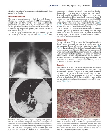
FIG 72.10 Radiographic Findings in Cryptogenic Organizing FIG 72.11 Histopathology of Cryptogenic Organizing Pneu-
Pneumonia. (A) Chest radiograph in a patient with cryptogenic monia. A photomicrograph of cryptogenic organizing pneumonia
organizing pneumonia shows bilateral patchy alveolar opacities shows intraalveolar fibroblast proliferation (arrows) and early
with a peripheral distribution in the setting of normal lung volumes. collagen production. In addition, thickening of the alveolar septa
(B) Chest computed tomography shows a dense right lower with a lymphoplasmacytic infiltrate consistent with cellular
lung consolidation with the presence of air bronchograms. nonspecific interstitial pneumonitis is present.