
Page 1009 - Clinical Immunology_ Principles and Practice ( PDFDrive )
P. 1009
CHaPter 72 Immunological Lung Diseases 973
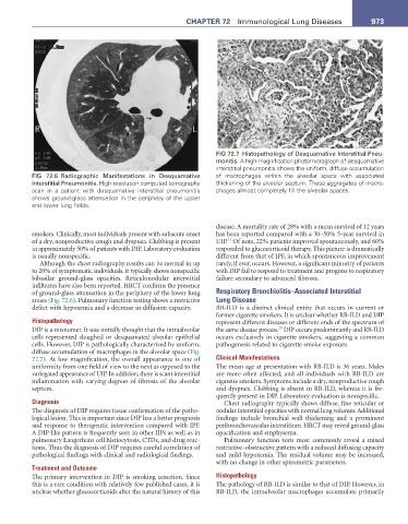
FIG 72.7 Histopathology of Desquamative Interstitial Pneu-
monitis. A high-magnification photomicrograph of desquamative
interstitial pneumonitis shows the uniform, diffuse accumulation
FIG 72.6 Radiographic Manifestations in Desquamative of macrophages within the alveolar space with associated
Interstitial Pneumonitis. High-resolution computed tomography thickening of the alveolar septum. These aggregates of macro-
scan in a patient with desquamative interstitial pneumonitis phages almost completely fill the alveolar spaces.
shows ground-glass attenuation in the periphery of the upper
and lower lung fields.
disease. A mortality rate of 28% with a mean survival of 12 years
smokers. Clinically, most individuals present with subacute onset has been reported compared with a 30–50% 5-year survival in
24
of a dry, nonproductive cough and dyspnea. Clubbing is present UIP. Of note, 22% patients improved spontaneously, and 60%
in approximately 50% of patients with DIP. Laboratory evaluation responded to glucocorticoid therapy. This picture is dramatically
is usually nonspecific. different from that of IPF, in which spontaneous improvement
Although the chest radiography results can be normal in up rarely, if ever, occurs. However, a significant minority of patients
to 20% of symptomatic individuals, it typically shows nonspecific with DIP fail to respond to treatment and progress to respiratory
bibasilar ground-glass opacities. Reticulonodular interstitial failure secondary to advanced fibrosis.
infiltrates have also been reported. HRCT confirms the presence
of ground-glass attenuation in the periphery of the lower lung Respiratory Bronchiolitis–Associated Interstitial
zones (Fig. 72.6). Pulmonary function testing shows a restrictive Lung Disease
defect with hypoxemia and a decrease in diffusion capacity. RB-ILD is a distinct clinical entity that occurs in current or
former cigarette smokers. It is unclear whether RB-ILD and DIP
Histopathology represent different diseases or different ends of the spectrum of
29
DIP is a misnomer. It was initially thought that the intraalveolar the same disease process. DIP occurs predominantly and RB-ILD
cells represented sloughed or desquamated alveolar epithelial occurs exclusively in cigarette smokers, suggesting a common
cells. However, DIP is pathologically characterized by uniform, pathogenesis related to cigarette smoke exposure.
diffuse accumulation of macrophages in the alveolar space (Fig.
72.7). At low magnification, the overall appearance is one of Clinical Manifestations
uniformity from one field of view to the next as opposed to the The mean age at presentation with RB-ILD is 36 years. Males
variegated appearance of UIP. In addition, there is scant interstitial are more often affected, and all individuals with RB-ILD are
inflammation with varying degrees of fibrosis of the alveolar cigarette smokers. Symptoms include a dry, nonproductive cough
septum. and dyspnea. Clubbing is absent in RB-ILD, whereas it is fre-
quently present in DIP. Laboratory evaluation is nonspecific.
Diagnosis Chest radiography typically shows diffuse, fine reticular or
The diagnosis of DIP requires tissue confirmation of the patho- nodular interstitial opacities with normal lung volumes. Additional
logical lesion. This is important since DIP has a better prognosis findings include bronchial wall thickening and a prominent
and response to therapeutic intervention compared with IPF. peribronchovascular interstitium. HRCT may reveal ground-glass
A DIP-like pattern is frequently seen in other IIPs as well as in opacification and emphysema.
pulmonary Langerhans cell histiocytosis, CTDs, and drug reac- Pulmonary function tests most commonly reveal a mixed
tions. Thus the diagnosis of DIP requires careful correlation of restrictive–obstructive pattern with a reduced diffusing capacity
pathological findings with clinical and radiological findings. and mild hypoxemia. The residual volume may be increased,
with no change in other spirometric parameters.
Treatment and Outcome
The primary intervention in DIP is smoking cessation. Since Histopathology
this is a rare condition with relatively few published cases, it is The pathology of RB-ILD is similar to that of DIP. However, in
unclear whether glucocorticoids alter the natural history of this RB-ILD, the intraalveolar macrophages accumulate primarily