
Page 1008 - Clinical Immunology_ Principles and Practice ( PDFDrive )
P. 1008
972 Part Seven Organ-Specific Inflammatory Disease
The history and physical findings in IPF are nonspecific.
However, extrapulmonary involvement does not occur; the pres-
ence of fever, arthralgias, myalgias, or pleuritis should suggest a
connective tissue disorder. Antinuclear antibodies (ANAs) and
rheumatoid factor are present in 10–20% of patients with IPF, but
titers greater than 1 : 320 should suggest an alternative diagnosis.
The majority of patients with IPF have abnormal chest
radiography results at the time of presentation. Basal peripheral
reticular opacities are the characteristic radiographic findings.
A confident diagnosis of IPF from HRCT of the lung requires
the presence of patchy, peripheral bibasal reticular abnormalities
23
with honeycombing. The presence of extensive ground-glass
opacities on HRCT should suggest an alternative diagnosis, such
as DIP, hypersensitivity pneumonitis, bronchiolitis obliterans
organizing pneumonia (BOOP), or NSIP.
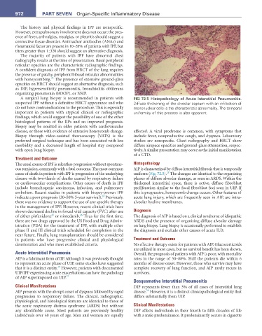
A surgical lung biopsy is recommended in patients with FIG 72.5 Histopathology of Acute Interstitial Pneumonitis.
suspected IPF without a definitive HRCT appearance and who Diffuse thickening of the alveolar septum with an infiltration of
do not have contraindications to the procedure. This is especially mononuclear cells is the characteristic abnormality. The temporal
important in patients with atypical clinical or radiographic uniformity of this process is also apparent.
findings, which could suggest the possibility of one of the other
histological patterns of the IIPs and an improved prognosis.
Biopsy may be omitted in older patients with cardiovascular
disease, or those with evidence of extensive honeycomb change. affected. A viral prodrome is common, with symptoms that
Biopsy through video-assisted thoracoscopy (VATS) is the include fever, nonproductive cough, and dyspnea. Laboratory
preferred surgical technique and has been associated with less studies are nonspecific. Chest radiography and HRCT show
morbidity and a decreased length of hospital stay compared diffuse airspace opacities and ground-glass attenuation, respec-
with open lung biopsy. tively. A similar presentation may occur as the initial manifestation
of a CTD.
Treatment and Outcome
The usual course of IPF is relentless progression without spontane- Histopathology
ous remission, commonly with a fatal outcome. The most common AIP is characterized by diffuse interstitial fibrosis that is temporally
28
cause of death in patients with IPF is progression of the underlying uniform (Fig. 72.5). The changes are identical to the organizing
disease with two-thirds of deaths caused by respiratory failure phases of diffuse alveolar damage, as seen in ARDS. Within the
or cardiovascular complications. Other causes of death in IPF thickened interstitial space, there is active, diffuse fibroblast
include bronchogenic carcinoma, infection, and pulmonary proliferation similar to the focal fibroblast foci seen in UIP. If
embolism. Recent studies in patients with biopsy-proven IPF this is progressive, honeycomb change occurs. Other features of
24
indicate a poor prognosis (30–50% 5-year survival). Previously, acute lung injury, which are frequently seen in AIP, are intra-
there was no evidence to support the use of any specific therapy alveolar hyaline membranes.
in the management of IPF. However, recent clinical trials have
shown decreased decline in forced vital capacity (FVC) after use Diagnosis
25
26
of either pirfenidone or nintedanib. Thus for the first time, The diagnosis of AIP is based on a clinical syndrome of idiopathic
there are two drugs approved by the US Food and Drug Admin- ARDS and the presence of organizing diffuse alveolar damage
istration (FDA) for the treatment of IPF, with multiple other on lung biopsy. Lung biopsy is occasionally performed to establish
phase II and III clinical trials scheduled for completion in the the diagnosis and exclude other causes of acute ILD.
near future. Finally, lung transplantation should be considered
in patients who have progressive clinical and physiological Treatment and Outcome
deterioration and who meet established criteria. No effective therapy exists for patients with AIP. Glucocorticoids
are utilized in most cases, but no survival benefit has been shown.
Acute Interstitial Pneumonia Overall, the prognosis of patients with AIP is poor, with mortality
AIP is a fulminant form of IIP. Although it was previously thought rates in the range of 50–88%. Half the patients die within 6
to represent an acute phase of UIP, some studies have suggested months of disease onset. However, those who survive may have
18
that it is a distinct entity. However, patients with documented complete recovery of lung function, and AIP rarely recurs in
UIP/IPF experiencing acute exacerbations can have the pathology survivors.
of AIP superimposed on UIP. 27
Desquamative Interstitial Pneumonitis
Clinical Manifestations DIP represents fewer than 3% of all cases of interstitial lung
29
AIP presents with the abrupt onset of dyspnea followed by rapid disease. However, it is a distinct clinicopathological entity that
progression to respiratory failure. The clinical, radiographic, differs substantially from UIP.
physiological, and histological features are identical to those of
the acute respiratory distress syndrome (ARDS) but without Clinical Manifestations
any identifiable cause. Most patients are previously healthy DIP affects individuals in their fourth to fifth decades of life
individuals over 40 years of age. Men and women are equally with a male predominance. It predominantly occurs in cigarette