
Page 268 - Clinical Immunology_ Principles and Practice ( PDFDrive )
P. 268
CHAPTER 17 Cytotoxic T Lymphocytes and Natural Killer Cells 249
cells are also a potent source of a diverse range of cytokines, THE CTL RESPONSE
including granulocyte macrophage–colony-stimulating factor
(GM-CSF), interleukin-5 (IL-5), IL-10, and IL-13 (Chapter 9). The CTL response to an acute infection consists of three phases:
The capacity to secrete a broad spectrum of cytokines provides NK first, the initial activation and proliferation of the CTL; second,
cells with a wide range of regulatory capabilities. CTLs and NK the contraction of effector populations; and third, the long-term
cells are capable of secreting a number of chemokines (Chapter maintenance of memory cells.
10), including chemokine ligand 3 (CCL3), CCL4, and CCL5.
These chemokines help recruit additional lymphocytes into the Initial Activation
immune reaction. Naïve T cells constantly circulate through secondary lymphoid
organs, where antigen encounter occurs. For a CTL response,
CYTOTOXIC T CELLS antigen is brought to the lymph node via the lymphatic system
by APC (Fig. 17.2). These APCs are typically DCs that mature
The Development and Tissue Distribution of CTLs after antigen acquisition in nonlymphoid tissues and migrate
CD8 T lymphocytes develop in the thymus, where they are selected to the lymph node. These antigens are recognized only when
for their ability to recognize nonself peptides in the context complexed to MHC molecules. APCs efficiently degrade self or
of MHC class I molecules (Chapter 8). Upon thymic export, foreign (pathogen-derived) proteins into shorter fragments
these cells acquire a quiescent state and are referred to as “naïve” (generally 8–10 amino acids in length) by the action of proteases
cells. Naïve CD8 T cells circulate between peripheral lymphoid in the cytosol. They are then transported into the lumen of the
organs, such as the spleen and lymph nodes, via the arterial endoplasmic reticulum, where they are loaded onto newly
and lymphatic systems. The tissue distribution of lymphocytes synthesized MHC class I molecules for presentation at the cell
is determined by targeting proteins, which can be divided into surface (Chapter 6). This then enables the APCs to communicate
5
three categories: selectins, chemokine receptors, and integrins. with the antigen-specific CD8 T cells via interactions between
Naïve and activated CD8 T cells display distinct sets of these the TCR and MHC molecules.
targeting proteins, allowing for the differential homing abili- In the lymph node, CTLs scan the APCs for the presence of
ties of these cells (Chapter 11). Naïve CD8 T cells express high antigenic peptides complexed with the MHC class I molecules,
levels of the lymph node homing receptor L-selectin (CD62L) a process termed immune surveillance. In the absence of a specific
and CCR7, a chemokine receptor that recognizes CCL19 and recognition by the TCR, the encounter is only transient, and the
CCL21, which are produced in the T-cell areas of secondary T cell continues on to another APC to repeat the process. If the
5
lymphoid organs (Table 17.1; Chapter 10). Here, naïve T cells MHC class I–peptide complex is bound by the TCR and initiates
interact with antigen-presenting cells (APCs), in particular signaling, a more lasting interaction occurs.
dendritic cells (DCs). If a naïve CD8 T cell does not encounter TCR activation promotes polarization of the T cell and forma-
5
6
its specific antigen, it leaves the lymph node. If, however, a tion of the “immunological synapse.” The immunological synapse
CD8 T cell encounters the correctly presented peptide–MHC is a highly structured body that functions to concentrate TCR
I complex, a dramatic change in its localization and homing signaling in a defined area. It is associated with the selective
properties ensues. These cells shut down their egress program recruitment of signaling molecules and exclusion of negative
and undergo multiple rounds of proliferation to become activated regulators. The synapse is stabilized by a ring of adhesion
CTLs. After the proliferative phase, the CTLs then reacquire molecules, including, for example, LFA-1, which binds to ICAM1
egress capacity and travel via the circulation to nonlymphoid on the APC (see Fig. 17.1). For a T cell to become fully active,
6
sites, where they tether to endothelial cells and extravasate costimulation through a second signaling pathway is required.
into tissue. This transmigration occurs in both inflamed and Many costimulators that have been identified share the common
noninflamed sites, such as skin, the gut, or the lung. Many of characteristic of being transmembrane receptors, often of the
the effector memory CTLs are retained in nonlymphoid tissues, TNFR superfamily, that bind transmembrane ligands on the
where they are poised to respond rapidly should the antigen be APC. The most important costimulator, CD28, binds the ligands
encountered again. The distinct types of memory T cells are CD80 (B7.1) and CD86 (B7.2), both of which are expressed on
discussed below. activated APCs. Costimulation results in the clonal expansion
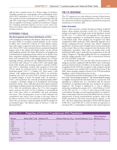
TABLE 17.1 Properties of Cytotoxic T Lymphocyte (CTL) Populations
Tissue-Resident
Marker Naïve CD8 T Cell Effector CTL Effector Memory (T EM ) Central Memory (T CM ) Memory (T RM )
CD69 – ++ – – ++
CD62L ++ – – ++ –
CD44 + +++ +++ +++ +++
CCR7 + – – + –
IL7R (CD127) ++ + + ++ +
IL2Rβ (CD122) + + ++ +++ +
Main tissue Lymph nodes, Lymph nodes, spleen, Nonlymphoid tissue (e.g., Lymph nodes, spleen, Nonlymphoid tissue
distribution spleen, blood blood, nonlymphoid lung, liver), spleen blood (e.g., lung, liver, skin)
tissue (e.g., lung, liver)
Cytotoxic function – +++ ++ – ++
IFN-γ – +++ +++ + +++