
Page 594 - Clinical Immunology_ Principles and Practice ( PDFDrive )
P. 594
572 ParT FIvE Allergic Diseases
malaise, loss of sense of smell (hyposmia), and cough. Of these,
nasal obstruction is the most common (81–95%), followed by
facial pain and pressure (70–85%), discolored nasal drainage
(51–83%), and hyposmia (61–69%). In contrast, symptoms in
children vary with age and require the parent or caregiver to
recognize them. Young children often present with a chronic
cough and irritability, rather than facial pain. Parents often also
report the child has halitosis and purulent nasal discharge.
Although it is more difficult to determine the prevalence of RS
in children because of overlapping symptoms with AR and viral
upper respiratory tract infections, its prevalence is inversely related
to age. 2
Diagnosis
The diagnosis of AR is based on a history of typical symptoms
and physical examination findings. Common symptoms include
postnasal drainage, sneezing, itchy nose and eyes, and clear
rhinorrhea. The frequency and effect of symptoms on sleep and
productivity should be assessed to classify the AR as intermittent
or persistent and mild or moderate–severe.
Patients suffering from AR can present with an “allergic shiner,”
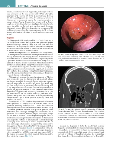
a darkening of the infraorbital skin resulting from chronic venous FIG 41.1 Nasal Polyposis. Seen on this nasal endoscopy are
pooling. In some children, wiping the front of the nose with the nasal polyps (NP) emanating from the sinus cavity into the nasal
back of the hand in an upward motion (the allergic salute) creates cavity between the septum (S) and the inferior turbinate (IT) of
a persistent horizontal crease across the nasal bridge that is a a patient with chronic rhinosinusitis.
hallmark of chronic anterior rhinorrhea. Bilateral conjunctivitis
may be present in patients along with ocular involvement.
On anterior rhinoscopy with a hand-held otoscope, engorged,
boggy, and pale inferior turbinates also suggest AR. In addition,
there is often a clear discharge coating the nasal cavity. Examina-
tion of the oropharynx often reveals cobblestoning of the mucosa,
a sign of chronic postnasal drip.
Although not necessary to make the diagnosis of AR, two
tests commonly used to demonstrate IgE-mediated allergic
reactions are immediate-hypersensitivity skin testing and measure-
ment of serum allergen–specific IgE levels. Skin prick tests
correlate well with the symptoms of allergic rhinitis and with
airway responsiveness to allergens, and measuring serum allergen-
specific IgE levels provides an in vitro means of supporting a
diagnosis of AR. Compared with skin prick testing, the in vitro
3
test is more specific but less sensitive and can be more expensive.
However, serum tests may be the only practical way of detecting
allergen-specific IgE in some patients, especially those with
urticaria and eczema.
The diagnosis of CRS requires the presence of at least two
major symptoms or one major and at least two minor clinical
symptoms persisting for longer than 12 weeks, in conjunction
with objective evidence of inflammation within the sinus cavity.
The major symptoms include facial pain or pressure, nasal FIG 41.2 Coronal Sinus Computed Tomography (CT) Image
obstruction, nasal drainage, and hyposmia or anosmia. Minor From a Patient With Chronic Rhinosinusitis. The maxillary
symptoms are headaches, halitosis, fatigue, dental pain, cough, sinuses (lateral to the nasal cavity) and ethmoid sinuses (medial
and ear pain or pressure. The most specific symptom for RS is to the orbital cavities) exhibit mucosal thickening and accumulation
4
discolored nasal discharge. Since most symptoms of rhinosinusitis of obstructed secretions consistent with inflammatory changes
are nonspecific, evidence of inflammation on nasal endoscopy within the paranasal sinuses.
or imaging is also necessary to make a diagnosis of CRS. On
nasal endoscopy, inflammation is suggested by edema and/or
drainage from the middle meatus; CRSwNP is diagnosed when To make the diagnosis of AFRS, the most widely accepted
nasal polyps are visualized (Fig. 41.1). Inflammation within the diagnostic criteria include five characteristics: nasal polyps; type
sinuses may not be apparent on nasal endoscopy: in patients I (immediate) hypersensitivity to fungi; radiographic imaging
with a strong history of CRS, computed tomography (CT) of consistent with AFRS; eosinophilic mucin with evidence of fungi;
the sinuses is necessary to detect mucosal thickening and/or and lack of evidence of fungal invasion into the surrounding
fluid within the sinuses (Fig. 41.2). sinus tissue. Type I hypersensitivity to fungi can be detected by