
Page 597 - Clinical Immunology_ Principles and Practice ( PDFDrive )
P. 597
CHaPTEr 41 Immunological Mechanisms of Airway Diseases and Pathways to Therapy 575
TABLE 41.1 Eosinophilic Lung Disorders
Proposed Immune
Disease Causative agent Mechanism
Loeffler syndrome Inhaled food, T cell–mediated
infection, or hypersensitivity
medication reaction
Drug rash with Drugs: Hypersensitivity reaction
eosinophilia and sulfonamides, to drug
systemic phenobarbital,
symptoms sulfasalazine,
(DRESS) carbamazepine,
syndrome and phenytoin
Parasitic infections Strongyloides spp., T-cell and B-cell clonal
Wuchereria activation in response
bancrofti, to parasite antigens
Brugia malayi and adjuvant factors
Allergic Aspergillus Immunoglobulin E (IgE)
bronchopulmonary and immune complex
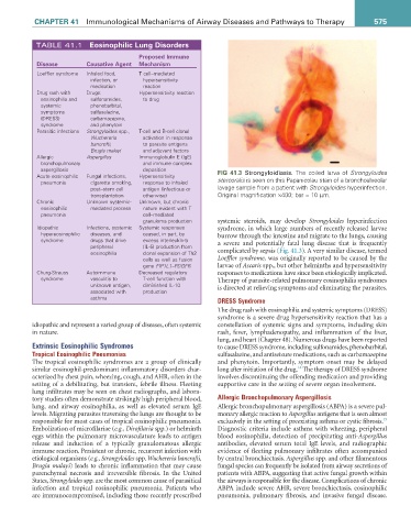
aspergillosis deposition FIG 41.3 Strongyloidiasis. The coiled larva of Strongyloides
Acute eosinophilic Fungal infections, Hypersensitivity
pneumonia cigarette smoking, response to inhaled stercoralis is seen on this Papanicolau stain of a bronchoalveolar
post–stem cell antigen (infectious or lavage sample from a patient with Strongyloides hyperinfection.
transplantation otherwise) Original magnification ×400; bar = 10 µm.
Chronic Unknown systemic- Unknown, but chronic
eosinophilic mediated process nature evident with T
pneumonia cell–mediated
granuloma production systemic steroids, may develop Strongyloides hyperinfection
Idiopathic Infections, systemic Systemic responses syndrome, in which large numbers of recently released larvae
hypereosinophilic diseases, and caused, in part, by burrow through the intestine and migrate to the lungs, causing
syndrome drugs that drive excess interleukin-5 a severe and potentially fatal lung disease that is frequently
peripheral (IL-5) production from
eosinophilia clonal expansion of Th2 complicated by sepsis (Fig. 41.3). A very similar disease, termed
cells as well as fusion Loeffler syndrome, was originally reported to be caused by the
gene FIP1L1–PDGFR larvae of Ascaris spp., but other helminths and hypersensitivity
Churg-Strauss Autoimmune Decreased regulatory responses to medications have since been etiologically implicated.
syndrome vasculitis to T-cell function with Therapy of parasite-related pulmonary eosinophilia syndromes
unknown antigen, diminished IL-10 is directed at relieving symptoms and eliminating the parasites.
associated with production
asthma DRESS Syndrome
The drug rash with eosinophilia and systemic symptoms (DRESS)
syndrome is a severe drug hypersensitivity reaction that has a
idiopathic and represent a varied group of diseases, often systemic constellation of systemic signs and symptoms, including skin
in nature. rash, fever, lymphadenopathy, and inflammation of the liver,
lung, and heart (Chapter 48). Numerous drugs have been reported
Extrinsic Eosinophilic Syndromes to cause DRESS syndrome, including sulfonamides, phenobarbital,
Tropical Eosinophilic Pneumonias sulfasalazine, and antiseizure medications, such as carbamazepine
The tropical eosinophilic syndromes are a group of clinically and phenytoin. Importantly, symptom onset may be delayed
14
similar eosinophil-predominant inflammatory disorders char- long after initiation of the drug. The therapy of DRESS syndrome
acterized by chest pain, wheezing, cough, and AHR, often in the involves discontinuing the offending medication and providing
setting of a debilitating, but transient, febrile illness. Fleeting supportive care in the setting of severe organ involvement.
lung infiltrates may be seen on chest radiographs, and labora-
tory studies often demonstrate strikingly high peripheral blood, Allergic Bronchopulmonary Aspergillosis
lung, and airway eosinophilia, as well as elevated serum IgE Allergic bronchopulmonary aspergillosis (ABPA) is a severe pul-
levels. Migrating parasites traversing the lungs are thought to be monary allergic reaction to Aspergillus antigens that is seen almost
15
responsible for most cases of tropical eosinophilic pneumonia. exclusively in the setting of preexisting asthma or cystic fibrosis.
Embolization of microfilariae (e.g., Dirofilaria spp.) or helminth Diagnostic criteria include asthma with wheezing, peripheral
eggs within the pulmonary microvasculature leads to antigen blood eosinophilia, detection of precipitating anti-Aspergillus
release and induction of a typically granulomatous allergic antibodies, elevated serum total IgE levels, and radiographic
immune reaction. Persistent or chronic, recurrent infection with evidence of fleeting pulmonary infiltrates often accompanied
etiological organisms (e.g., Strongyloides spp. Wuchereria bancrofti, by central bronchiectasis. Aspergillus spp. and other filamentous
Brugia malayi) leads to chronic inflammation that may cause fungal species can frequently be isolated from airway secretions of
parenchymal necrosis and irreversible fibrosis. In the United patients with ABPA, suggesting that active fungal growth within
States, Strongyloides spp. are the most common cause of parasitical the airways is responsible for the disease. Complications of chronic
infection and tropical eosinophilic pneumonia. Patients who ABPA include severe AHR, severe bronchiectasis, eosinophilic
are immunocompromised, including those recently prescribed pneumonia, pulmonary fibrosis, and invasive fungal disease.