
Page 63 - Clinical Immunology_ Principles and Practice ( PDFDrive )
P. 63
48 ParT ONE Principles of Immune Response
Toll-like receptor increased expression of MHC molecules along with expression
IL-1/18 receptor of costimulatory molecules (e.g., B7–1, B7–2, and IL-12p70)
results in subsequent development of adaptive immune responses.
Leucine-rich repeats (LRR) IL-12p70 stimulates IFN-γ production by T cells, which further
ligand binding augments the microbicidal activities of phagocytes. Stimulation
of TLR3, -7, -8, and -9 elicits the production of proinflammatory
cytokines, as well as type 1 IFNs, which play a crucial role in
Toll-IL-1 receptor innate antiviral immunity and also influence adaptive immune
Domain (TIR) responses.
Engagement of TLRs activates complex signaling pathways
MyD88 that have been characterized through biochemical analyses and
in gene-targeted mice. 51-53 TLR, IL-1R, and IL-18R share similar
signaling pathways (Fig. 3.5). Upon ligand binding, the cyto-
plasmic adaptor protein MyD88 is recruited to the TIR domain
Downstreaming signaling of the receptor for all TLRs, except TLR3. Recruitment of MyD88
leads to recruitment of IL-1 receptor associated kinase-4 (IRAK-4),
likely through death domain interactions. IRAK-4 activation
Transcriptional activation leads to recruitment and activation of IRAK-1 and IRAK-2. In
Nucleus monocytes, IRAK-M is also recruited to this complex and
functions as a negative regulator of signaling. Both IRAK-1 and
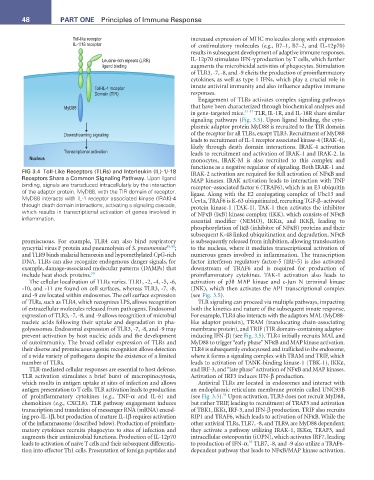
FIG 3.4 Toll-Like Receptors (TLRs) and Interleukin (IL)-1/-18 IRAK-2 activation are required for full activation of NFκB and
Receptors Share a Common Signaling Pathway. Upon ligand MAP kinases. IRAK activation leads to interaction with TNF
binding, signals are transduced intracellularly by the interaction receptor–associated factor 6 (TRAF6), which is an E3 ubiquitin
of the adaptor protein, MyD88, with the TIR domain of receptor. ligase. Along with the E2 conjugating complex of Ubc13 and
MyD88 interacts with IL-1 receptor associated kinase (IRAK)-4 Uev1a, TRAF6 is K-63 ubiquitinated, recruiting TGF-β–activated
through death domain interactions, activating a signaling cascade, protein kinase-1 (TAK-1). TAK-1 then activates the inhibitor
which results in transcriptional activation of genes involved in of NFκB (IκB) kinase complex (IKK), which consists of NFκB
inflammation. essential modifier (NEMO), IKKα, and IKKβ, leading to
phosphorylation of IκB (inhibitor of NFκB) proteins and their
subsequent K-48 linked ubiquitination and degradation. NFκB
promiscuous. For example, TLR4 can also bind respiratory is subsequently released from inhibition, allowing translocation
syncytial virus F protein and pneumolysin of S. pneumoniae 48,49 ; to the nucleus, where it mediates transcriptional activation of
and TLR9 binds malarial hemozoin and hypomethylated CpG-rich numerous genes involved in inflammation. The transcription
DNA. TLRs can also recognize endogenous danger signals, for factor interferon regulatory factor-5 (IRF-5) is also activated
example, damage-associated molecular patterns (DAMPs) that downstream of TRAF6 and is required for production of
include heat shock proteins. 50 proinflammatory cytokines. TAK-1 activation also leads to
The cellular localization of TLRs varies. TLR1, -2, -4, -5, -6, activation of p38 MAP kinase and c-Jun N terminal kinase
-10, and -11 are found on cell surfaces, whereas TLR3, -7, -8, (JNK), which then activates the AP1 transcriptional complex
and -9 are located within endosomes. The cell surface expression (see Fig. 3.5).
of TLRs, such as TLR4, which recognizes LPS, allows recognition TLR signaling can proceed via multiple pathways, impacting
of extracellular molecules released from pathogens. Endosomal both the kinetics and nature of the subsequent innate response.
expression of TLR3, -7, -8, and -9 allows recognition of microbial For example, TLR4 also interacts with the adaptors MAL (MyD88-
nucleic acids following their uptake and degradation in pha- like adaptor protein), TRAM (translocating chain-associating
golysosomes. Endosomal expression of TLR3, -7, -8, and -9 may membrane protein), and TRIF (TIR domain–containing adaptor-
prevent activation by host nucleic acids and the development inducing IFN-β) (see Fig. 3.5). TLR4 initially recruits MAL and
of autoimmunity. The broad cellular expression of TLRs and MyD88 to trigger “early phase” NFκB and MAP kinase activation.
their diverse and promiscuous agonist recognition allows detection TLR4 is subsequently endocytosed and trafficked to the endosome,
of a wide variety of pathogens despite the existence of a limited where it forms a signaling complex with TRAM and TRIF, which
number of TLRs. leads to activation of TANK-binding kinase-1 (TBK-1), IKKε,
TLR-mediated cellular responses are essential to host defense. and IRF-3, and “late phase” activation of NFκB and MAP kinases.
TLR activation stimulates a brief burst of macropinocytosis, Activation of IRF3 induces IFN-β production.
which results in antigen uptake at sites of infection and allows Antiviral TLRs are located in endosomes and interact with
antigen presentation to T cells. TLR activation leads to production an endoplasmic reticulum membrane protein called UNC93B
54
of proinflammatory cytokines (e.g., TNF-α and IL-6) and (see Fig. 3.5). Upon activation, TLR3 does not recruit MyD88,
chemokines (e.g., CXCL8). TLR pathway engagement induces but rather TRIF, leading to recruitment of TRAF3 and activation
transcription and translation of messenger RNA (mRNA) encod- of TBK1, IKKι, IRF-3, and IFN-β production. TRIF also recruits
ing pro-IL-1β, but production of mature IL-1β requires activation RIP1 and TRAF6, which leads to activation of NFκB. While the
of the inflammasome (described below). Production of proinflam- other antiviral TLRs, TLR7, -8, and TLR9, are MyD88 dependent;
matory cytokines recruits phagocytes to sites of infection and they activate a pathway utilizing IRAK-1, IKKα, TRAF3, and
augments their antimicrobial functions. Production of IL-12p70 intracellular osteopontin (iOPN), which activates IRF7, leading
55
leads to activation of naïve T cells and their subsequent differentia- to production of IFN-α. TLR7, -8, and -9 also utilize a TRAF6-
tion into effector Th1 cells. Presentation of foreign peptides and dependent pathway that leads to NFκB/MAP kinase activation.