
Page 684 - Clinical Immunology_ Principles and Practice ( PDFDrive )
P. 684
CHaPTEr 48 Drug Hypersensitivity 657
Keratinocyte
ICAM-1 MCH II
Keratinocyte
cell necrosis
Perforin
Hydropic
degeneration
LFA-1 TCR Granzyme B
Eosinophils
Mononuclear
cell infiltrate
A B Drug-specific CD4 + T cell
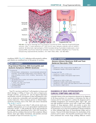
FiG 48.5 (A) Typical histology of a maculopapular exanthema (MPE). Note the focal keratinocyte
necrosis, often in close apposition of T cells (which have cytotoxic potential and are perforin-
positive). (B) Schematic representation of CD4-mediated killing of activated keratinocytes, which
express MHC class II and intercellular adhesion molecule 1 (ICAM1). (Modified from Pichler WJ.
Delayed drug hypersensitivity reactions. Ann Intern Med. 2003; 139: 683–693.)
exanthema (MPE) (Fig. 48.5), infections with nematodes, asthma, KEY CONCEPT
and rhinitis are considered part of this group of reactions.
Stevens-Johnson Syndrome (SJS) and Toxic
KEY CONCEPT Epidermal Necrolysis (TEN)
Drug-Induced Reactions With Eosinophilia and SJS
Systemic Symptoms (DRESS) Syndrome • Detachment <10% of body surface area (BSA), plus
• Widespread macules or flat atypical target lesions
• Drug-induced
• Anticonvulsants, antidepressants, non-antimicrobial sulfones/sul- Overlap SJS-TEN
fonamides, nonsteroidal antiinflammatory drugs (NSAIDs), angio- • Detachment 10–30% of BSA, plus
tensin-converting enzyme (ACE) inhibitors, beta-blockers, and • Widespread macules or flat atypical target lesions
antibiotics
• Systemic disorder TEN
• Hematological abnormalities (eosinophilia, atypical lymphocytes)
• Multiorgan involvement • Detachment of >30% of BSA, plus
• kidney, liver, heart, lung, thyroid • Widespread macules or flat atypical target lesions
• Hypogammaglobulinemia • Detachment >10% of BSA with large epidermal sheets and without
• Lymphadenopathy any macules or target lesions
• Skin involvement
Type IVc reactions result from T-cell migration to tissues and DIAGNOSIS OF DRUG HYPERSENSITIVITY:
direct damage or killing of tissue cells, such as hepatocytes, CLINICAL SYMPTOMS AND HISTORY
through perforin-, granzyme B– and Fas-ligand–dependent
mechanisms, with the recruitment of other inflammatory cells, A detailed history is the most important element in the diagnosis
such as monocytes, eosinophils, and neutrophil polymorpho- of all drug reactions; it is necessary to determine the type of
nuclear leukocytes. These reactions present as delayed hyper- reaction and the test required to confirm the diagnosis and to
sensitivity reactions, such as SJS, TEN, and contact dermatitis establish management and treatment plans. Type I IgE- and
(Fig. 48.4B, C and D). non–IgE-mediated reactions occur with a rapid onset, within
Type IVd reactions occur when T cells activate sterile neu- minutes of drug exposure, which can present as pure skin
trophilic inflammation, as in acute generalized exanthematous manifestations or as involvement of other organs. Sudden onset
pustulosis (AGEP). T cells producing CXCL8 and granulocyte of hypotension can be the first manifestation during anesthesia
macrophage–colony-stimulating factor (GM-CSF) recruit leu- or when infusing high doses of chemotherapy drugs, such as
kocytes, and the cytokines then prevent their apoptosis. Behçet taxanes. Tryptase levels help determine the mechanism of the
disease, linear IgA bullous dermatosis, and pustular psoriasis reaction and its extent. The basophil activation test (BAT) has
are examples of type IVd reactions (Fig. 48.6). been performed for several drugs yielding different results;