
Page 71 - Clinical Immunology_ Principles and Practice ( PDFDrive )
P. 71
56 Part one Principles of Immune Response
combined immune deficiency (SCID) in the case of T cells strand distribution. The two additional strands, which encode
(Chapter 35). framework region 2 (FR2), are used to steady the interaction
between heterodimeric V domains, allowing them to create a
IMMUNOGLOBULINS AND TCR STRUCTURES stable antigen-binding site. 2
Although each Ig or TCR chain contains only one amino-
The Ig Domain, the Basic IgSF Building Block terminal V Ig domain, the number of carboxy-terminal C domains
Igs consist of two heavy (H) and two light (L) chains (Fig. 4.1). varies. Ig H chains contain between three and four C domains,
The L chain can be either a κ or a λ chain. TCRs consist of either whereas both Ig L chains and all four TCR chains contain only
an αβ or a γδ heterodimer. Each component chain contains two one C domain each. IgH chains with three C domains tend to
or more IgSF domains, each of which consists of two sandwiched include a spacer hinge region between the first (C H 1) and second
β pleated sheets “pinned” together by a disulfide bridge between (C H 2) domains. Each V or C domain consists of approximately
1
two conserved cysteine residues. Considerable variability is 110–130 amino acids, averaging 12 000–13 000 kilodaltons (kDa).
allowed to the amino acids that populate the external surface of A typical L chain will mass approximately 25 kDa, whereas a
the IgSF domain and the loops that link the β strands. These three C domain Cγ H chain with its hinge and tail will mass
solvent exposed surfaces offer multiple targets for docking with approximately 55 kDa.
other molecules.
Two types of IgSF domains, “constant” (C) and “variable” Idiotypes and Isotypes
(V), are used in Igs and TCRs (see Fig. 4.1). C-type domains, Immunization of heterologous species with monoclonal antibodies
which are the most compact, have seven antiparallel strands (mAbs; or a restricted set of Igs) has shown that Igs and TCRs
distributed as three strands in the first sheet and four strands contain both common and individual antigenic determinants.
in the second. Side chains positioned to lie between the two Individual determinant(s), termed idiotype(s), are contained
strands tend to be nonpolar in nature, creating a hydrophobic within V domains. Common determinants, termed isotypes, are
core of considerable stability. V-type domains add two additional specific for the constant portion of the antibody and allow
antiparallel strands to the first sheet, creating a five-strand–four- grouping of Igs and TCRs into recognized classes. Each class
defines an individual type of C domain. Determinants common
to subsets of individuals within a species, yet differing between
other members of that species, are termed allotypes and define
D C domain inherited polymorphisms that result from allelic forms of the
genes. 3
C E The V Domain
NH 2
B Early comparisons of the primary sequences of V domains
identified three hypervariable intervals, termed complementarity-
F determining regions (CDRs), situated between four framework
A
regions of stable sequence (Fig. 4.2). The current definition of
these regions integrates sequence diversity with three-dimensional
4
G structure. The international ImMunoGeneTics information
system (IMGT) maintains an extremely useful website (http://
A
COOH www.imgt.org), which contains a large database of Ig and TCR
sequences and numerous software tools for their analyses.
V domain
Antigen Recognition and Fab
D
Studies of Ig structure were facilitated by the use of papain and
C˝ E pepsin to fragment IgG molecules. Papain digests IgG into two
C´ B antigen-binding fragments (Fab) and a single crystallizable (or
constant) fragment (Fc). Pepsin splits IgG into an Fc fragment
F C and a single dimeric F(ab’) 2 that can cross-link as well as bind
A antigens. Fab contains one complete L chain in its entirety and
G the V and C H 1 portion of one H chain (see Fig. 4.2). Fab can
NH 2 be further divided into a variable fragment (Fv) composed of
COOH the V H and V L domains and a constant fragment (Fb) composed
of the C L and C H 1 domains. Single Fv fragments can be genetically
engineered to recapitulate the monovalent antigen-binding
B characteristics of the original, parent antibody. The extracellular
5
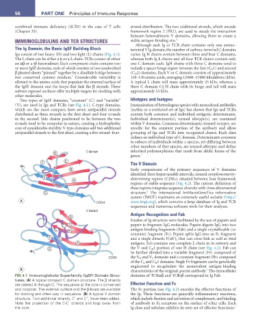
FIG 4.1 Immunoglobulin Superfamily (IgSF) Domain Struc- domains of TCRαβ and TCRγδ correspond to Ig Fab.
tures. (a) A typical compact C domain structure. The β strands
are labeled A through G. The sequence at the core is conserved Effector Function and Fc
and nonpolar. The external surface and the β-loops are available The Fc portion (see Fig. 4.2) encodes the effector functions of
for docking and often vary in sequence. (B) A typical V domain the Ig. These functions are generally inflammatory reactions,
structure. Two additional strands, C’ and C”, have been added. which include fixation and activation of complement, and binding
Note the projection of the C-C’ strands and loop away from of antibody to Fc receptors on the surface of other cells. Each
6
the core. Ig class and subclass exhibits its own set of effector functions.