
Page 964 - Clinical Immunology_ Principles and Practice ( PDFDrive )
P. 964
CHaPter 68 Immunological Renal Diseases 931
For patients with “pure” class V lupus MN and nephrotic-range
proteinuria, both the ACR and the EULAR/ERA-EDTA have
recommended oral prednisone and MMF. A prospective controlled
trial showed that both IVCY and CSA were more effective than
steroids alone in inducing remission of proteinuria in lupus MN
but that relapse of nephrotic syndrome occurred significantly
more often in the CSA group than in the IVCY treatment group.
Scleroderma (Systemic Sclerosis) Renal Disease
KeY COnCePtS
Nephropathies of Selected Connective
Tissue Diseases
• Scleroderma renal crisis: Predominantly renal vasculopathy; moderate
to severe (high renin) hypertension with progressive renal failure—
treated with angiotensin-converting enzyme inhibitors (ACEIs); additional
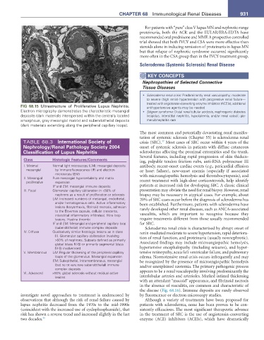
FIG 68.15 Ultrastructure of Proliferative Lupus Nephritis. antihypertensive agents may be needed
Electron micrography demonstrates the characteristic mesangial • Sjögren syndrome: Distal renal tubular acidosis, nephrogenic diabetes
deposits (dark materials interspersed within the centrally located insipidus, interstitial nephritis, hypokalemia, and/or renal calculi; glo-
amorphous, gray mesangial matrix) and subendothelial deposits merulonephritis rare
(dark materials extending along the peripheral capillary loops).
The most common and potentially devastating renal manifes-
tation of systemic sclerosis (Chapter 55) is scleroderma renal
TABLE 68.3 International Society of crisis (SRC). Most cases of SRC occur within 4 years of the
47
nephrology/renal Pathology Society 2004 onset of systemic sclerosis in patients with diffuse cutaneous
Classification of Lupus nephritis scleroderma affecting the proximal extremities and the trunk.
Several features, including rapid progression of skin thicken-
Class Histologic Features/Comments ing, palpable tendon friction rubs, anti-RNA polymerase III
I. Minimal Normal light microscopy (LM); mesangial deposits antibody, recent-onset cardiac events (e.g., pericardial effusion
mesangial by immunofluorescence (IF) and electron or heart failure), new-onset anemia (especially if associated
microscopy (EM) with microangiopathic hemolysis and thrombocytopenia), and
II. Mesangial Pure mesangial hypercellularity and matrix
proliferative expansion recent treatment with high-dose corticosteroids, help identify
IF and EM: mesangial immune deposits patients at increased risk for developing SRC. A classic clinical
III. Focal Glomerular capillary obliteration in <50% of presentation may obviate the need for renal biopsy. However, renal
nephrons as a result of proliferation or sclerosis biopsy may be necessary in atypical cases. For example, about
LM: Increased numbers of mesangial, endothelial, 20% of SRC cases occur before the diagnosis of scleroderma has
and/or hematogenous cells. Active inflammatory been established. Furthermore, patients with scleroderma have
lesions (karyorrhexis, fibrinoid necrosis, adhesion rarely developed other renal diseases, such as ANCA-associated
to the Bowman capsule, cellular crescents,
interstitial inflammatory infiltrates). Wire loop vasculitis, which are important to recognize because they
lesions. Hyaline thrombi require treatments different from those usually recommended
IF and EM: Mesangial and peripheral capillary loop for SRC.
(subendothelial) immune complex deposits Scleroderma renal crisis is characterized by abrupt onset of
IV. Diffuse Qualitatively similar histologic lesions as in class renin-mediated moderate to severe hypertension, rapid deteriora-
III. Glomerular capillary obliteration involving tion of renal function, and proteinuria (usually nonnephrotic).
>50% of nephrons. Subsets defined as primarily
global (class IV-G) or primarily segmental (class Associated findings may include microangiopathic hemolysis,
IV-S) involvement hypertensive encephalopathy (including seizures), and hyper-
V. Membranous LM: Regular thickening of the peripheral capillary tensive retinopathy, acute left ventricular failure, and pulmonary
loops of the glomerulus. Mesangial expansion edema. Normotensive renal crisis occurs infrequently and may
EM: Subepithelial, intramembranous, mesangial be recognized by the presence of microangiopathic hemolysis
(but no or very rare subendothelial) immune and/or unexplained azotemia. The primary pathogenic process
complex deposits
VI. Advanced >90% global sclerosis without residual active appears to be a renal vasculopathy involving predominantly the
lesions interlobular arteries and arterioles. Marked intimal thickening
with an attendant “mucoid” appearance, and fibrinoid necrosis
in the absence of vasculitis, are common and characteristic of
the disease (Fig. 68.16). Immune deposits are rarely observed
investigate novel approaches to treatment is underscored by by fluorescence or electron microscopy studies.
observations that although the risk of renal failure caused by Although a variety of treatments have been proposed for
lupus nephritis decreased from the 1970s to the mid-1990s patients with scleroderma, none has been proven to be con-
(coincident with the increased use of cyclophosphamide), that sistently efficacious. The most significant therapeutic advance
risk has shown a reverse trend and increased slightly in the last in the treatment of SRC is the use of angiotensin-converting
two decades. 46 enzyme (ACE) inhibitors (ACEIs), which have dramatically