
Page 1268 - Hall et al (2015) Principles of Critical Care-McGraw-Hill
P. 1268
CHAPTER 93: Oncologic Emergencies 875
51
NEUROLOGIC EMERGENCIES failed to show improved outcomes with this practice. Levetiracetam and
■ STATUS EPILEPTICUS for SE, have also been used effectively. 52,53 After the administration of
valproic acid, while not approved by the Food and Drug Administration
Status epilepticus (SE) can be a life-threatening emergency when unrec- any antiepileptic drug, clinical and EEG evaluation should be performed
to determine if further seizure activity persists.
ognized and left untreated. SE is defined as either a persistent seizure Refractory status epilepticus (RSE) is defined as seizures that persist
for >30 minutes or repeated seizures with no recovery of consciousness after adding a second line of medication therapy. 45,46 In RSE, only 7.3%
between each episode. Importantly, any seizure activity that continues of patients respond to administration of a second drug, and 2% to a
for >5 minutes should be treated as SE. 41,42 The incidence of SE in the third drug. Even though there are reports of survival after prolonged
51
United States is 18.3 to 41 episodes per 100,000 patients/year and about periods of SE, the prognosis is poor. 41,42,44 Refractory SE requires further
7% of seizures progress to SE. In the cancer population, about 13% of workup including reevaluation of administered doses of antiepilep-
41
patients experience seizures at some point during the course of their tic drugs (AED), discontinuation of medications that could decrease
disease; 50% of these seizures are secondary to brain metastasis or pri- the therapeutic levels of AED, and further imaging such as magnetic
mary brain tumors. Nevertheless, the prevalence of SE is no higher in resonance imaging (MRI), position emission tomography (PET), and
43
cancer patients when compared to the general population. 44 single-photon emission computed tomography (SPECT) to rule out
The causes for SE are the same in the general population and in patients 41
with cancer. The most common causes are noncompliance to medica- unidentified structural pathologies. After further workup has been
performed, medication-induced coma is the next step in the treatment
tions (29%), alcohol intoxication or withdrawal (26%), CNS infection of RSE. In these cases, continuous EEG monitoring is the most reliable
(8%), stroke (6%), tumors (6%), trauma (6%), and anoxic encephalopathy method for the evaluation of responsiveness to treatment.
(6%). In cancer patients, it is important to rule out malignancy-related Benzodiazepine infusion is used for the initial treatment of RSE.
44
causes of SE (Table 93-1). Poor prognostic factors are presence of anoxia, However, their efficacy becomes reduced with prolonged use. Surveys
42
old age, multiple comorbidities, and brain tumors. 41,42 performed in Europe and North America report that barbiturates are the
Initial management of patients with SE should focus on stabilizing the first drug of choice for RSE among neurologists and neurointensivists.
47
patient’s airway and cardiovascular status. Laboratory workup should be Secondary effects of barbiturates include cardiovascular depression,
performed to determine any possible metabolic or toxic causes as well aplastic anemia, and liver dysfunction. Propofol has also been used
41
as brain imaging to determine structural lesions. In patients with poor successfully for the treatment of RSE. There are no studies suggesting
mental status or coma in whom nonconvulsive SE is suspected, an elec- any benefits of propofol over benzodiazepines; however, barbiturates
troencephalogram (EEG) should be obtained. Lumbar puncture should have shown higher success rates and lower breakthrough seizures when
be considered if an infectious cause is suspected. Supportive treatment compared to benzodiazepines and propofol. Ketamine, lidocaine, and
54
for SE includes avoiding hyperthermia, hypoxia, hypotension, hypergly- inhaled anesthetics have also been used for RSE. If there is no response
41
cemia, hyperventilation, and electrolyte imbalance. 45,46 Treatment and after a single medication infusion, a combination should be considered,
stabilization of the patient should not be delayed by diagnostic tests and being cautious of possible potentiating side effects. Favorable outcomes
procedures since multiple studies have shown that delaying treatment of have been reported in patients who received simultaneous midazolam
SE causes brain injury, and increases morbidity and mortality. 45,47 Delaying and propofol infusions. Multiple studies have proven the efficiency
55
treatment of SE can decrease the response to treatment from 80% to 30% of newer drugs such as lacosamide and sec-butyl-propylacetamide
and therefore increase the risk for refractory SE. Moreover, institutional (a derivative of valproic acid) in SE. 56,57 Hypothermia, described in case
45
protocols for treatment of SE have shown to improve outcomes. 42,45 reports and studied in animal models, appears to also have an encourag-
Initial management of SE should be with intermittent boluses of 58,59
benzodiazepines. 47,48 There are four double-blind randomized controlled ing role in the treatment of SE. However, further studies are required
before any of these newer treatment options become standard of care.
trials comparing diazepam or midazolam with lorazepam. 48-51 While Titration of infusions should be considered after 24 to 48 hours of
initial studies did not show any preference for one benzodiazepine over no seizure activity on EEG. 42,45 Slow titration, while continuing other
the other, Alldredge et al reported that the use of lorazepam led to earlier AEDs, should be performed with careful observation for epileptiform
termination of SE when compared to diazepam. Addition of hydanto- activity on EEG or clinical evaluation. If all of these measures are inef-
49
ins (phenytoin, fosphenytoin) to initial boluses of benzodiazepines is fective, surgical intervention, electroconvulsive therapy, and transcranial
recommended due to the decreased efficacy of benzodiazepines after magnetic stimulation should be considered. 41,45,46
30 minutes of sustained seizure activity although large studies have
42
■ MALIGNANT SPINAL CORD COMPRESSION
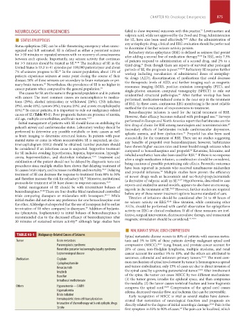
TABLE 93-1 Malignancy-Related Causes of Seizures Spinal metastatic disease occurs in 40% of patients with osseous metas-
Tumor Brain metastasis tasis and 5% to 10% of these patients develop malignant spinal cord
Paraneoplastic syndrome compression (MSCC). 60,61 Lung, breast, and prostate cancer account for
Reversible posterior leukoencephalopathy syndrome 20% of cases; non-Hodgkin lymphoma, multiple myeloma, and renal
Leptomeningeal disease cancer account for another 5% to 10%, and the others are attributed to
Medication Cisplatin sarcomas, colorectal and unknown primary tumors. 62-65 The most com-
Cyclophosphamide mon mechanism of spinal involvement by tumor is hematogenous spread
Bevacizumab and tumor embolization; only 15% of cases are due to direct invasion of
60,62
Imatinib the spinal canal by a growing paravertebral tumor. After involvement
Busulfan of the spine, the tumor can cause MSCC by two different mechanisms:
Intrathecal methotrexate (1) the tumor grows, invades the epidural space, and then compresses
the medulla; (2) the tumor causes vertebral fracture and bone fragments
Others Hyponatremia—SIADH compress the spinal cord. 60,64 Compression of the spinal cord causes
Hypercalcemia edema, decreased vascular flow, and ischemia that can be irreversible.
Brain radiation Early recognition of MSCC is vital as several studies have demon-
Hematopoietic stem cell transplantation strated that restoration of neurological function and prognosis are
Interaction of chemotherapy with anti epileptic drugs directly related to the degree of initial neurologic damage. 62,66 Pain is the
Stroke first symptom in 83% to 90% of cases. The pain can be localized, which
64
section07.indd 875 1/21/2015 7:42:58 AM