
Page 1273 - Hall et al (2015) Principles of Critical Care-McGraw-Hill
P. 1273
880 PART 7: Hematologic and Oncologic Disorders
embolism in this population. 127,128 However, bleeding complications are are mesotheliomas. Pericardial effusions due to malignancy are
102
high, and current data do not suggest improved outcomes when using associated with a poor prognosis; only 45% of patients survive at
routine anticoagulation. 123 6 months decreasing to 10% to 26% at 1 year. 132,133 The worst outcomes
The use of intravascular stents for SVCS treatment has increased have been associated with patients with lung cancer, especially adeno-
significantly since the early 1980s. Stenting improves symptoms in the carcinoma, in whom survival is less than 3 months after pericardial effu-
129
first 24 to 72 hours, making it the treatment of choice for patients with sions are diagnosed. 132,134 On the contrary, patients with lymphoma have
severe SVCS. 126,129 Multiple studies have shown stenting to be efficient, a better prognosis with survival of up to 3 years. 134,135 Other prognostic
safe, and cost-effective. 126,129,130 Complications such as stent migration, factors associated with higher mortality are positive fluid cytology, no
pericardial tamponade, and PE have been reported in less than 8% of response to chemotherapy, and advanced malignancy. 132,136
cases. Moreover, stenosis of stents has been reported to be less than The most common symptoms associated with cardiac tamponade are
129
7%. 129,130 Prolonged use of anticoagulants to avoid stent thrombosis is dyspnea, chest discomfort, and chest pain. Beck triad, consisting of pulsus
still controversial and some authors have observed that dipyridamole paradoxus, distant heart sounds, and jugular venous distention (JVD),
may be enough to avoid this complication. 130 was first described in the 1930s. However, JVD, hypotension, and muffled
As described earlier, only in cases of cerebral edema and airway heart sounds have been only described in 54%, 28%, and 22% of cases,
compromise should treatment for SVCS be initiated urgently and respectively. Chest x-ray reveals an enlarged cardiac silhouette in about
137
without a diagnosis. Even after stent placement, final treatment for 70% of cases. Electrocardiographic signs consistent with pericardial
137
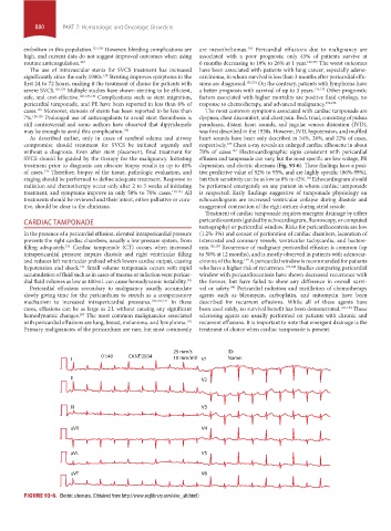
SVCS should be guided by the therapy for the malignancy. Initiating effusion and tamponade can vary, but the most specific are low voltage, PR
treatment prior to diagnosis can obscure biopsy results in up to 48% depression, and electric alternans (Fig. 93-6). These findings have a posi-
of cases. Therefore, biopsy of the tissue, pathologic evaluation, and tive predictive value of 92% to 95%, and are highly specific (86%-99%),
123
staging should be performed to define adequate treatment. Response to but their sensitivity can be as low as 8% to 42%. Echocardiogram should
138
radiation and chemotherapy occur only after 2 to 3 weeks of initiating be performed emergently on any patient in whom cardiac tamponade
treatment, and symptoms improve in only 50% to 70% cases. 120,123 All is suspected. Early findings suggestive of tamponade physiology on
treatments should be reviewed and their intent, either palliative or cura- echocardiogram are increased ventricular collapse during diastole and
tive, should be clear to the clinicians. exaggerated contraction of the right atrium during atrial systole.
Treatment of cardiac tamponade requires emergent drainage by either
CARDIAC TAMPONADE pericardiocentesis (guided by echocardiogram, fluoroscopy, or computed
tomography) or pericardial window. Risks for pericardiocentesis are low
In the presence of a pericardial effusion, elevated intrapericardial pressure (1.2%-3%) and consist of perforation of cardiac chambers, laceration of
prevents the right cardiac chambers, usually a low pressure system, from intercostal and coronary vessels, ventricular tachycardia, and bactere-
filling adequately. Cardiac tamponade (CT) occurs when increased mia. 136,139 Recurrence of malignant pericardial effusion is common (up
131
intrapericardial pressure impairs diastole and right ventricular filling to 50% at 12 months), and is mostly observed in patients with adenocar-
and reduces left ventricular preload which lowers cardiac output, causing cinoma of the lung. A pericardial window is recommended for patients
139
hypotension and shock. Small volume tamponade occurs with rapid who have a higher risk of recurrence. 139,140 Studies comparing pericardial
131
accumulation of fluid such as in cases of trauma or infection were pericar- window with pericardiocentesis have shown decreased recurrence with
dial fluid volumes as low as 400 mL can cause hemodynamic instability. 102 the former, but have failed to show any difference in overall survi-
Pericardial effusions secondary to malignancy usually accumulate val or safety. Pericardial radiation and instillation of chemotherapy
140
slowly giving time for the pericardium to stretch as a compensatory agents such as bleomycin, carboplatin, and mitomycin have been
mechanism to increased intrapericardial pressures. 102,103,131 In these described for recurrent effusions. While all of these agents have
cases, effusions can be as large as 2 L without causing any significant been used safely, no survival benefit has been demonstrated. 141-144 These
hemodynamic changes. The most common malignancies associated sclerosing agents are usually performed on patients with chronic and
103
with pericardial effusions are lung, breast, melanoma, and lymphoma. recurrent effusions. It is important to note that emergent drainage is the
102
Primary malignancies of the pericardium are rare, but most commonly treatment of choice when cardiac tamponade is present.
25 mm/s ID:
01:40 CANT:2834
I 10 mm/mV V1 Name:
II V2
III V3
aVR V4
aVL V5
aVF V6
FIGURE 93-6. Electric alternans. (Obtained from http://www.ecglibrary.com/elec_alt.html)
section07.indd 880 1/21/2015 7:43:02 AM