
Page 1351 - Hall et al (2015) Principles of Critical Care-McGraw-Hill
P. 1351
924 PART 8: Renal and Metabolic Disorders
as an equivalent loss of isotonic fluid (all of which must come from the ability is diminished (renal dysfunction or advanced age), higher urine
smaller ECF compartment). Thus the importance of changes in weight volumes are required to maintain adequate solute excretion. Since such
should be assessed relative to changes in serum sodium concentration. conditions are more the rule than the exception, it seems more appro-
The cardinal signs of ECF volume depletion are changes in hemody- priate to expect solute retention at urine outputs below the more typical
namic parameters, jugular venous pressure, and in the skin. An ortho- ICU monitoring target of 0.5 to 1 mL/kg per hour (840-1680 mL/d). In
static increase in pulse of 15 beats per minute or a decrease in diastolic the RIFLE, AKIN, and KDIGO classification systems, oliguria (defined
blood pressure of 10 mm Hg can detect losses of 5% of the ECF volume. as a urine output <0.5 mL/kg per minute) persisting for 6 hours or
A postural increase in pulse (supine to standing) of at least 30 beats longer is defined as AKI, and more severe and/or persistent oliguria is
per minute is 96% specific for clinically significant volume depletion, classified as higher stage AKI, irrespective of serum creatinine trends. Of
whereas systolic pressure may fall 20 mm Hg upon standing in 10% course, urine output targets must be sufficient to control fluid balance as
of normal individuals and in up to 30% of patients less than age 65. well as solute excretion, so higher urine output values may be required
90
The inability of a patient to stand because of severe lightheadedness is for patients with large obligate fluid intakes.
a relatively specific sign of hypovolemia. Skin changes that accompany AKI may be classified as anuric (urine output <100 mL/d), oliguric
volume depletion include cool, mottled extremities, dry mucous mem- (urine output <400 mL/d), or nonoliguric (urine output >400 mL/d).
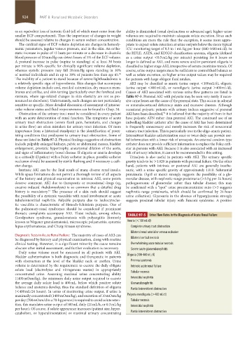
branes and axillae, and skin tenting (particularly over the forehead and Causes of AKI associated with various urine flow patterns are listed in
sternum, where age-related changes in skin elasticity are not as pro- Table 97-5. Prerenal AKI with polyuria may be seen very rarely if exces-
nounced as elsewhere). Unfortunately, such changes are not particularly sive urine losses are the cause of the prerenal state. This occurs in adrenal
sensitive or specific. More detailed discussion of assessment of intravas- or mineralocorticoid deficiency states and excessive diuresis. Although
cular volume status and fluid responsiveness can be found in Chap. 34. occasional polyuric patients with urinary indices suggestive of prerenal
Obstruction of the urinary tract must be considered in every patient AKI have been described, it is believed that the majority of them in fact
92
with an acute deterioration of renal function. The symptoms of acute have polyuric ATN rather than prerenal AKI. The continued use of an
urinary tract obstruction (severe flank pain, hematuria, and changes indwelling bladder catheter after the cause of AKI has been determined
in urine flow) are often mistaken for urinary tract infection. Of more is frequently unnecessary and merely increases the risk of nosocomial
importance from a historical standpoint is the identification of preex- urinary tract infection. This is particularly true in the oligo-anuric patient.
isting conditions that predispose to urinary tract obstruction. Some of Intermittent bladder catheterization once or twice daily can provide use-
these are listed in Table 97-2. Physical findings suggestive of obstruction ful information with a lower risk of urosepsis. An external condom-type
include palpably enlarged kidneys, pelvic or abdominal masses, bladder catheter does not provide sufficient information to replace the Foley cath-
enlargement, prostatic hypertrophy, aneurysmal dilation of the aorta, eter in persons with AKI. Because it is also associated with an increased
and signs of inflammatory bowel disease. If oliguria or anuria develops risk of urinary infection, it cannot be recommended in this setting.
in a critically ill patient with a Foley catheter in place, possible catheter Urinalysis is also useful in patients with AKI. The urinary specific
occlusion should be assessed by sterile flushing and if necessary a cath- gravity tends to be >1.020 in patients with prerenal failure. On the other
eter change. hand, patients with intrinsic or postrenal AKI are generally isosthe-
Intrinsic AKI can be the final result of many diverse renal insults. nuric, with a urine specific gravity of approximately 1.010. Substantial
While space limitations do not permit a thorough review of all aspects proteinuria (3 g/d or more) strongly suggests the possibility of a glo-
of the history and physical examination in intrinsic AKI, some points merular disease, with nephrotic-range proteinuria (>3.5 g per 24 hours)
deserve comment. AKI due to therapeutic or recreational drugs (eg, pathognomonic of glomerular rather than tubular disease; this may
cocaine-induced rhabdomyolysis) is so common that a detailed drug be confirmed with a “spot” urine protein:creatinine ratio (>3 suggests
91
history is mandatory. The presence of a skin rash should suggest nephrotic-range proteinuria, which should be confirmed by 24-hour
the possibility of a systemic vasculitis with renal involvement or acute urine collection). Glycosuria in the absence of hyperglycemia strongly
tubulointerstitial nephritis. Palpable purpura due to leukocytoclas- suggests proximal tubular injury with Fanconi syndrome. A positive
tic vasculitis is characteristic of Henoch-Schönlein purpura. One of
the pulmonary-renal syndromes should be considered if prominent
thoracic complaints accompany AKI. These include, among others, TABLE 97-5 Urine Flow Rates in the Diagnosis of Acute Renal Failure
Goodpasture syndrome, granulomatosis with polyangiitis (formerly
known as Wegener granulomatosis), microscopic polyarteritis, systemic Anuria (<100 mL/d)
lupus erythematosus, and Churg-Strauss syndrome. Complete urinary tract obstruction
Bilateral renal arterial or venous occlusion
Diagnostic Tests in Acute Renal Failure: The majority of cases of AKI can
be diagnosed by history and physical examination, along with routine Bilateral cortical necrosis
clinical testing. However, in a significant minority the cause remains Overwhelming acute tubular necrosis
obscure after initial assessment, and further evaluation is necessary. Severe acute glomerulonephritis
Daily urine volume must be measured in all patients with AKI.
Bladder catheterization is both diagnostic and therapeutic in patients Oliguria (100-400 mL/d)
with obstruction at the level of the bladder neck or urethra. Urine Prerenal azotemia
volume is determined by the requirement to excrete the daily obligate Intrinsic acute renal failure
solute load (electrolytes and nitrogenous wastes) in appropriately
concentrated urine. Assuming maximal urine concentrating ability Tubular necrosis
(1400 mOsm/kg), the minimum daily urine output required to excrete Interstitial nephritis
the average daily solute load is 400 mL, below which positive solute Glomerulonephritis
balance and azotemia develop, thus the standard definition of oliguria Partial intermittent obstruction
(<400 mL/24 hours). In terms of monitoring urine output, if urine is
maximally concentrated (1400 mOsm/kg), and excretion of 10 mOsm/kg Polyuria nonoliguria (>400 mL/d)
per day (700 mOsm/d in a 70-kg person) is required to avoid solute reten- Tubular necrosis
tion, this mandates urine output of 500 mL daily (21 mL/h, or 0.3 mL/kg Interstitial nephritis
per hour). Of course, if solute appearance increases (patient size, hyper-
catabolism, or hyperalimentation) or maximal urinary concentrating Partial intermittent obstruction
section08.indd 924 1/14/2015 8:27:56 AM