
Page 1352 - Hall et al (2015) Principles of Critical Care-McGraw-Hill
P. 1352
CHAPTER 97: Acute Kidney Injury 925
reaction for blood in the urine is consistent with acute glomerular or concentrations, respectively. The normal FE is 50% to 65%, reflect-
UN
tubular injury, urinary tract infection, or nephrolithiasis. If blood is ing reabsorption of approximately 50% of filtered urea in the proximal
present on dipstick but not microscopically, or if the findings are dispro- tubule; urea reabsorption is trivial in the thick ascending limb and distal
portionate (eg, 4+ blood on dipstick with rare erythrocytes on micros- convoluted tubule. Hypovolemia results in increased urea absorption,
copy), a pigment nephropathy (hemoglobinuria or myoglobinuria) decreased urea clearance, and thus a lower FE . Loop and thiazide
95
UN
should be considered. The urine sediment is usually unremarkable in diuretics, which act at the thick ascending limb and distal convoluted
prerenal and postrenal azotemia, except for occasional hyaline casts. tubule, do not interfere directly with urea reabsorption and should not
In postrenal AKI due to stones, blood and crystals can be seen. Intrinsic alter FE . However, proximal tubule diuretics and osmotic diuresis
UN
AKI is often associated with a characteristic (or even diagnostic) urine decrease proximal reabsorption of urea and may produce an inappro-
sediment. A careful microscopic examination frequently can distinguish priately high FE . Carvounis and colleagues prospectively evaluated
UN
between GN, AIN, ATN, and TIN. Erythrocyte casts, often accompanied 102 hospitalized patients with AKI. Patients were divided into three
87
by proteinuria and numerous erythrocytes and leukocytes, are pathog- groups: 50 were deemed prerenal; 27 were deemed prerenal with diuret-
nomonic of GN. Detection of large numbers of leukocytes, leukocyte ics given up to the day of consultation (details were not provided as to
casts, and eosinophils in uninfected urine strongly suggests the diagnosis whether diuretics were given 1 or 23 hours prior to the urine sample);
of drug-induced AIN. ATN is suggested by findings including muddy and 25 were diagnosed with ATN. Patients with AIN, GN, and obstruc-
brown granular casts, free renal tubular cells, and tubular cell casts. tive nephropathy were excluded. Fe was <1%, as expected, in 92% of
Na
Several measurements of urine composition have been suggested as group 1 patients, but in only 48% of the prerenal patients treated with
ways to differentiate between prerenal azotemia and intrinsic AKI in diuretics. In contrast, 90% of the group 1 patients and 89% of those given
the oliguric patient. Urine electrolytes are most useful in this regard, diuretics had a FE <35%. The ATN patients evidenced a mean FE
93
UN
UN
especially the fractional excretion of sodium (FE ), calculated as of 59%. A FE <35% had 85% sensitivity, 92% specificity, 99% positive
UN
Na predictive value, and 75% negative predictive value for a prerenal state.
U × P × 100
FE (%) = Na Cr (97-1) In this study, the urine:plasma creatinine ratio also performed better
P × U
Na
Na
Na Cr than FE to distinguish prerenal azotemia from ATN. One other urine
chemistry test may be useful in hyperuricemic patients with AKI and
where U and P are urine and plasma sodium concentrations, respec- possible urate nephropathy (tumor lysis syndrome and hypovolemia
Na
Na
tively, and U and P are urine and plasma creatinine concentrations, with hyperuricemia and acid urine): urine uric acid:urine creatinine
Cr
Cr
respectively. Values of FE <0.01 (1%) in oliguric patients suggest ratios >1.1 are consistent with acute urate nephropathy. 96
Na
avid tubular sodium reclamation and prerenal azotemia with function- Urine microscopy may also be useful in distinguishing between intrin-
ing renal tubules, whereas values >0.03 (3%) suggest tubular injury. sic AKI and prerenal AKI, and has the advantage of being widely available
The FE is less useful in patients who are not oliguric. Contrary to and inexpensive. However, expertise is required in interpretation. In a
94
Na
common belief, however, it may be useful in diuretic-treated patients. study of 267 patients with AKI, a urine sediment scoring system based on
Although an elevated value may be a result of ATN or the effects of the presence of casts and renal tubular epithelial cells was highly predictive
the diuretic, a low level in the face of diuretic therapy strongly suggests of AKI. In patients with a high pretest probability of ATN, the presence of
volume depletion and prerenal AKI. Some causes of AKI presenting any casts or renal tubular epithelial cells resulted in a positive predictive
with a low FE are listed in Table 97-6. A low U (<10 mEq/L) as value of 100% and negative predictive value of 41% for ATN. In patients
Na
Na
an isolated measurement is often used as evidence of a prerenal state. with a low pretest probability of ATN, the lack of casts or renal tubular
However, this measurement depends exquisitely on the state of water cells was associated with a negative predictive value for ATN of 91%. 97
balance in addition to sodium balance. It cannot be said that it is any Although the diagnosis and staging of AKI is currently based on
easier to use than FE , since an independent evaluation of water balance indices of kidney function (acute changes in serum creatinine or cys-
Na
must be made to interpret it. Therefore, it is not recommended as an tatin, BUN, or the development of oliguria), the search for earlier and
isolated measurement in the routine evaluation of AKI. more sensitive biomarkers of renal tubular damage (in search of a “renal
Most other urinary diagnostic indices do not show any clear-cut troponin”) has been an active area of investigations in recent years.
98
superiority over FE in distinguishing prerenal azotemia from ATN; These tools have begun to undergo clinical evaluation in a number
Na
however, they are independently useful in the assessment of tubular of health systems internationally. Early studies suggest that such tools
function. Recent data suggest urinary fractional excretion of urea (FE ) may not only help to diagnose AKI with evolving ATN early (facilitat-
UN
is superior to FE to distinguish prerenal azotemia from ATN, particu- ing potentially successful clinical trials of new therapies), but will also
Na
larly in diuretic-treated patients. FE is calculated as help to distinguish reversible, prerenal functional AKI from AKI with
UN
99
U × P × 100 structural kidney damage (ATN), and to predict which cases of AKI are
FE (%) = UN Cr (97-2) likely to progress, require RRT, and have worse clinical outcomes. 98-101
UN BUN × U
Cr Finally, it is hoped that emerging tools in development for “real-time”
monitoring of GFR (as opposed to following serum creatinine changes
where U and BUN are urine and serum urea nitrogen concentra-
UN
tions, respectively, and U and P are urine and plasma creatinine hours-to-days after renal function is lost) will also help to dynamically
Cr Cr assess kidney function in critically ill patients. 102
Several radiographic studies are useful in the evaluation of patients
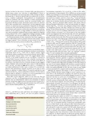
TABLE 97-6 Causes of Acute Renal Failure With Low Fractional Excretion of Sodium with AKI. Plain films of the abdomen can assess kidney size, detect
>90% of renal stones, and detect skeletal abnormalities of secondary
Prerenal azotemia
hyperparathyroidism, which imply established CKD rather than AKI.
Nonoliguric acute tubular necrosis In our view, the potential hazards of intravenous pyelography make
Acute glomerulonephritis this test of little benefit in the work-up of AKI. Renal ultrasound is a
sensitive and specific method for detecting hydronephrosis. It is prob-
Acute obstruction (early)
ably indicated in nearly every patient with AKI unless obstruction can
Acute interstitial nephritis be proven more quickly in another manner (eg, by bladder catheter-
Contrast nephropathy ization in a patient with symptoms of bladder neck obstruction) or if
Nontraumatic rhabdomyolysis some diagnosis other than obstruction is made with certainty early in
the evaluation. If clinical suspicion of obstruction persists despite an
Uric acid nephropathy
apparently negative ultrasound, retrograde pyelography is the definitive
section08.indd 925 1/14/2015 8:27:56 AM