
Page 1353 - Hall et al (2015) Principles of Critical Care-McGraw-Hill
P. 1353
926 PART 8: Renal and Metabolic Disorders
diagnostic maneuver. Computed tomography of the kidneys does not result of tumor lysis syndrome. This is noted most commonly in patients
103
have any advantages over ultrasound and retrograde pyelography in the with germ cell tumors or hematologic malignancies because of their
detection of obstruction, but may help clarify its cause (eg, detection of rapid turnover. The tumor lysis syndrome is due to the toxic effects
104
obstructing ureteral stones, or compressing tumor). Radionuclide scans of intracellular constituents, which cause AKI, hyperkalemia, hyper-
of the kidneys may be useful in the evaluation of patients with suspected phosphatemia, hypocalcemia, and hyperuricemia. The most common
vascular accidents of the kidneys. pathogenetic mechanism involved is acute urate nephropathy. When
serum uric acid levels exceed about 20 mg/dL, the risk of AKI is high.
CLINICAL ACUTE RENAL FAILURE SYNDROMES The urinary ratio of uric acid to creatinine is >1. Uric acid crystals may
be seen in the urinary sediment. Synergistic factors in the development
There are a number of diseases that commonly lead to AKI. In many of the acute nephropathy include volume depletion and acidosis. The
cases of AKI in the ICU, prerenal azotemia combines with exposure to usual pathophysiology of urate nephropathy is intratubular deposition
nephrotoxins (endogenous or exogenous) to cause ATN. However, the of urate crystals, causing intrarenal obstruction. Less commonly,
105
suspicion of other causes such as obstruction or specific parenchymal ureteral obstruction may be seen.
lesions is increased in certain patient populations. Once oliguria occurs, diuretics are seldom useful in restoring urine
■ MALIGNANCY AND ACUTE RENAL FAILURE flow. Allopurinol and alkaline diuresis are effective measures when insti-
tuted prophylactically, but do not reverse established urate nephropathy.
106
AKI is seen in patients with malignancy and could be caused by malig- Rasburicase, a recombinant form of urate oxidase, has been shown to
nancy itself or its treatment with chemotherapeutic agents (Table 97-7). be effective in reducing plasma uric acid levels within 4 hours after the
107
The full differential diagnosis of AKI must be considered in cancer first dose ; urate oxidase converts uric acid to allantoin, which is more
patients, including common causes such as prerenal azotemia, drug soluble than uric acid. Severe hyperphosphatemia (levels >20 mg/dL)
108
nephrotoxicity, and obstructive uropathy, as well as rarer causes such can be associated with tumor lysis and subsequent AKI. The
as tumor infiltration of the kidneys and radiation nephritis. Urinary pathogenesis of the renal dysfunction can be either intratubular crys-
tract obstruction leading to AKI is commonly seen in patients with tallization of phosphate or metastatic calcification in the renal paren-
malignancy. Ureteral obstruction is seen in patients with abdominal and chyma. It is important to make the distinction between this entity and
pelvic metastasis. AKI is also caused by intratubular obstruction. This is urate nephropathy, because urinary alkalinization promotes calcium
usually seen in patients with acute uric acid nephropathy and/or calcium phosphate crystallization. If hyperuricemia is prevented or controlled by
phosphate crystallization as part of tumor lysis syndrome, intratubular volume expansion, allopurinol, or uricase, it may be preferable to avoid
obstruction by light chains in patients with multiple myeloma, and in or stop urinary alkalinization if severe hyperphosphatemia develops,
patients who received high-dose methotrexate. to avoid hyperphosphatemia AKI and the risk of tetany (hypocalcemia
A variety of chemotherapeutic agents are potentially nephrotoxic, combined with metabolic alkalosis). Renal replacement therapy in
including cisplatin, nitrosoureas, and methotrexate, as discussed in tumor lysis syndrome is indicated in patients in whom the resolution
Chap. 952. In addition, mitomycin is known to cause hemolytic uremic of AKI is unlikely, and is also indicated in patients with life-threatening
syndrome thus leading to renal failure. fluid and electrolyte abnormalities. Normalization of uric acid and phos-
If large numbers of malignant cells suddenly die, as in spontaneous phorus levels is required for recovery of renal function. Hemodialysis is
necrosis or successful chemotherapy of large tumors, AKI can occur as a far more effective in the clearance of uric acid compared to peritoneal
dialysis. Continuous renal replacement therapies (CRRT) have been
104
used in the management of tumor lysis syndrome. The solute clear-
ances vary depending on the specific prescription. In these patients
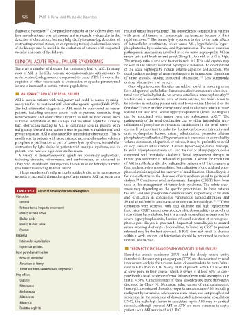
TABLE 97-7 Causes of Renal Dysfunction in Malignancy
the uric acid and phosphorus clearances were, respectively, 45 mL/min
Obstruction and 47 mL/min in continuous venovenous hemodiafiltration and
Ureteral 39 and 40 mL/min in continuous arteriovenous hemodialysis. 109,110 These
clearances were achieved with high dialysate and high replacement
Retroperitoneal lymphatic involvement
fluid rates. CRRT cannot correct electrolyte abnormalities as rapidly as
Primary ureteral tumor intermittent hemodialysis, but it is a much more effective treatment for
Bladder neck severe hyperphosphatemia, because rebound elevation of serum phos-
Primary bladder tumor phorus post dialysis is prevented. Sequential hemodialysis to control
severe evolving electrolyte abnormalities, followed by CRRT to prevent
Prostate rebound may be the best approach. If RRT does not result in diuresis
Intrarenal within a week, ureteral catheterization should be performed to exclude
Intratubular crystallization ureteral obstruction.
Light-chain proteins ■ THROMBOTIC MICROANGIOPATHY AND ACUTE RENAL FAILURE
Renal parenchymal invasion
Hemolytic uremic syndrome (HUS) and the closely related entity
Renal cell carcinoma thrombotic thrombocytopenic purpura (TTP) are characterized by renal
Metastases to kidneys involvement early in their course. Renal disease tends to be more fulmi-
nant in HUS than in TTP. Nearly 100% of patients with HUS have AKI
Tumor infiltration (leukemias and lymphomas)
at some point in their course (which is severe in at least 60%) as com-
Drug effects pared with a total incidence of renal failure of even mild severity in TTP
Cisplatin that is <50%. Clinical features of these disorders are more thoroughly
discussed in Chap. 91. Numerous other causes of microangiopathic
Nitrosoureas hemolytic anemia and thrombocytopenia can also cause AKI, including
Methotrexate malignant hypertension, scleroderma renal crisis, and antiphospholipid
Mithramycin syndrome. In the syndrome of disseminated intravascular coagulation
Mitomycin (DIC), the pathologic lesion in associated septic AKI may be cortical
necrosis, although prerenal AKI or ATN are more common in septic
Radiation nephritis patients with AKI associated with DIC.
section08.indd 926 1/14/2015 8:27:56 AM