
Page 1397 - Hall et al (2015) Principles of Critical Care-McGraw-Hill
P. 1397
970 PART 8: Renal and Metabolic Disorders
The treatment of metabolic acidosis requires treatment of an underlying
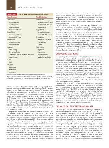
TABLE 100-1 Potential Clinical Effects of Metabolic Acid-Base Disorders
disease process and not, strictly speaking, of the acid-base disorder. If
Metabolic Acidosis Metabolic Alkalosis the patient developed a lactate acidosis following a seizure, this lactic
Cardiovascular Cardiovascular acidosis would resolve rapidly once the liver metabolized the lactate.
Indeed, treatment of acid-base disturbances can lead to severe overshoot
Decreased inotropy Increased inotropy (Ca entry)
2+
alkalosis or acidosis.
Conduction defects Altered coronary blood flow a Finally, the liver is perhaps the most important abdominal organ
Arterial vasodilation Digoxin toxicity involved in the regulation of acid-base balance. 10,11 Hepatic glutami-
nogenesis is important for systemic acid-base balance and is tightly
Venous vasoconstriction Oxygen delivery
controlled by mechanisms sensitive to plasma [H ] and is stimulated
+
Oxygen delivery Increased oxy-Hb affinity by acidosis. Nitrogen metabolism by the liver can produce urea,
12
+
Decreased oxy-Hb binding Increased 2,3-DPG (delayed) glutamine, or NH . Normally, the liver does not release more than a
4
very small amount of NH but incorporates this nitrogen into either
+
Decreased 2,3-DPG (late) Neuromuscular 4
urea or glutamine. However, the production of urea or glutamine has
Neuromuscular Neuromuscular excitability significantly different effects at the level of the kidney. This is so because
Respiratory depression Encephalopathy glutamine is used by the kidney to generate NH and facilitate the
+
4
−
Decreased sensorium Seizures excretion of Cl . Thus the production of glutamine can be seen as hav-
ing an alkalinizing effect on plasma pH because of the way in which the
Metabolism Metabolic effect kidney uses it. In humans, the liver is also the only organ that synthesizes
Protein wasting Hypokalemia albumin, the major component of A .
tot
Bone demineralization Hypocalcemia
Catecholamine, PTH, and aldosterone stimulation Hypophosphatemia CRYSTALLOID SOLUTIONS
+
Insulin resistance Impaired enzyme function Manipulating [H ] in the blood is intellectually easy once one under-
stands the importance of SID but certainly is not of proven benefit.
GI effect
When administered to patients, equimolar concentrations of Na and
+
Emesis Cl (such as in saline solutions) will increase the [Cl ] more rapidly than
−
−
+
−
+
Electrolytes the [Na ] because [Na ] is normally much greater than [Cl ]. When this
occurs, SID will decrease, and [H ] will increase. In a test tube, lactated
+
Hyperkalemia
Ringer solution will behave just like saline because lactate is a strong ion.
Hypercalcemia However, in humans, lactate metabolism is rapid even under conditions
Hyperuricemia of relatively severe hepatic dysfunction. If the liver is functioning and
+
a Animal studies have shown both increased and decreased coronary artery blood flow. can metabolize lactate, then the unbalanced Na will increase the SID
and result in alkalemia. Conversely, if lactate-containing solutions are
Adapted with permission from Kellum JA. Diagnosis and treatment of acid-base disorders. In: Grenvik A, administered quickly (as in replacement fluid for hemofiltration) and
Shoemaker PK, Ayers S, Holbrook, eds. Textbook of Critical Care. Philadelphia, PA: Saunders; 1999.
hepatic function is impaired, acidosis will develop, just as in the case of
saline loading, because SID is lowered.
Normal saline (0.9% NaCl) is often blamed for causing a “dilutional”
acidosis, but all that is occurring is that [Na ] is relatively unchanged as
+
different portions of the gastrointestinal tract. Cl is pumped into the the [Cl ] rises, leading to a decreased SID and hyperchloremic acidosis.
−
−
stomach, reducing SID in the stomach and increasing the [H ] (decreas- Adding 75 mEq/L of [NaHCO3] to 0.45% saline (77 mEq/L Na and
+
+
ing the pH) and, at the same time, causing the alkaline tide in the blood 77 mEq/L Cl ) will create an isotonic solution that contains half the
−
(increasing SID) that occurs at the beginning of a meal when gastric [Cl ] (a strong anion) with twice the [Na ] (strong cation). This solu-
−
+
acid secretion is maximal. The alkaline tide refers to the Cl -depleted tion has a higher SID than normal saline or lactated Ringer solution
−
plasma that leaves the stomach. The elevated SID leads to a decrease in and favors alkalemia. Mixing 150 mEq NaHCO in 1 L of sterile water
[H ] (increase in pH). Cl is reabsorbed in the duodenum, and plasma increases the SID further and creates an even more potent alkalizing
3
−
+
[H ] or pH is restored. Given the combination of Cl secretion into the fluid. Again, it is worth emphasizing the need to treat the underlying
+
−
stomach and Cl reabsorption in the duodenum, a net balance occurs, disorder and not just “correct” the acid-basis disorder.
−
and plasma [H ] or pH is not affected. However, if gastric secretions are
+
removed from the patient by nasogastric (NG) suction or by vomiting, THE ANION GAP AND THE STRONG ION GAP
Cl cannot be reabsorbed, and SID will increase. Increased SID will lead
−
to a metabolic alkalosis. The anion gap (AG) was popularized over 30 years ago. Traditionally,
The pancreas secretes fluid into the small intestine, which has a SID it is calculated from the equation [(Na ) + (K )] − [(Cl ) + (HCO )];
+
+
−
−
3
that is much higher than plasma and very low in [Cl ]. The Cl -rich K is often omitted because its plasma concentration is so tightly con-
−
+
−
plasma leaving the pancreas counteracts the alkaline tide along with trolled that there is little variation. However, this is a mistake for two
Cl reabsorption in the duodenum. Large amounts of pancreatic fluid reasons. First, a 2- to 3-mEq difference in the AG may be clinically
−
loss will lead to a decrease in plasma SID and an associated acidosis. relevant in some scenarios, and second, techniques used to correct the
At the other end of the gastrointestinal (GI) tract, in the large intestine, AG for abnormalities in [A ] require a full accounting of other ions.
tot
most of the Cl already has been removed in the small intestine, so the The difference in the gap is made up largely by albumin and, to a lesser
−
only strong ions present are Na and K . If large amounts of these strong extent, phosphate. Other anions, such as sulfate and lactate, normally
+
+
ions are lost with diarrhea fluid, then the plasma SID will decrease, and contribute less than 2 mEq of negative charge, similar in fact to the
acidosis will result. During ischemia to the intestinal tract, significant amount of positive charge contributed by ionized calcium and ionized
amounts of lactate can be produced. At physiologic pH, lactate acts as a magnesium. Thus these ions tend to offset each other. Many medical
strong anion and decreases SID, leading to a metabolic acidosis. There is textbooks still report a normal range for the AG of about 12 to 16 mEq
some evidence that the gut may modulate systemic acidosis in experimen- (when K is considered). This value, however, is based on older assay
+
tal endotoxemia by removing anions from the plasma. However, the methods that were less sensitive for Cl ; the expected AG using modern
−
9
full capacity of the GI tract to affect acid-base balance is not known. analyzers is closer to 8 to 10 mEq. However, many critically ill patients
section08.indd 970 1/14/2015 8:28:26 AM