
Page 1394 - Hall et al (2015) Principles of Critical Care-McGraw-Hill
P. 1394
CHAPTER 99: Electrolyte Disorders in Critical Care 967
hypokalemia, hypocalcemia, and hyponatremia. Symptoms due to hypo- excretion of magnesium. A 24-hour urine magnesium level less than
magnesemia become more common as serum magnesium falls below 20 mg is consistent with an intact renal response to hypomagnesemia
1.2 mg/dL (Table 99-17). and implicates decreased intake or extrarenal losses as the cause of
280
Neuromuscular Effects Neuromuscular irritability is a common sign of mag- hypomagnesemia. A 24-hour urinary magnesium level greater than
nesium depletion. Patients can develop Trousseau and Chvostek signs 24 mg indicates renal magnesium wasting. The fractional excretion of
despite normal ionized calcium. Severe depletion can cause weakness, magnesium (FeMg) allows assessment of the renal handling of mag-
fatigue, vertical nystagmus, tetany, and seizures. Reversible blindness nesium on a single urine specimen. A cutoff of 4% correctly separates
281
due to magnesium deficiency has been reported. 282 patients with renal magnesium wasting (FeMg >4%) from patients
287
Metabolic Effects Hypokalemia is commonly associated with hypomagne- with decreased magnesium absorption (FeMg <4%) (Eq. 99-7).
semia. One series reported it to occur in 40% of patients with hypo-
magnesemia. The reverse is also true; 60% of patients with hypokalemia FeMg = 100 × sCr× uMg
are hypomagnesemic. One explanation for this phenomenon is that (.07 × sMg ×) uCr
261
many of the etiologies of hypomagnesemia (diuretics, alcoholism,
and diarrhea, among others) also result in hypokalemia. In addition, Equation 99-7. The fractional resorption of magnesium differentiates between
hypomagnesemia causes renal potassium wasting. The mechanism for magnesium-avid and magnesium-wasting states. An FeMg greater than 4% in the presence of
potassium wasting is multifactorial. Decreased intracellular magne- hypomagnesemia indicates abnormal renal magnesium wasting. sCr, serum creatinine; sMg,
sium slows adenosine triphosphate (ATP) production, which decreases serum magnesium; uCr, urine creatinine; uMg, urine magnesium.
Na-K-ATPase activity, resulting in the loss of intracellular potassium. Clinical sequelae of altered magnesium content are more dependent
In the TALH and cortical collecting duct, the loss of ATP increases the on tissue magnesium levels than blood magnesium concentration.
number of potassium channels on the apical membrane. Intracellular Isolated tissue magnesium depletion (normomagnesemic magnesium
70
potassium flows down its concentration gradient into the tubule and is deficiency) may be a cause of refractory hypokalemia or hypocalcemia,
lost in the urine. especially in those at high risk of magnesium deficiency. One method
284
Hypocalcemia has been reported in 12% to 50% of patients with to infer the tissue magnesium level in patients with normal serum mag-
hypomagnesemia. 278,283 Hypomagnesemia suppresses the release of nesium is a physiologic test that measures the renal response to a magne-
PTH and causes end-organ resistance to PTH. The hypocalcemia is sium load. An 800 mg infusion of magnesium is given over 8 hours, and
refractory to calcium supplementation until the magnesium deficit is a 24-hour urine is collected starting from the initiation of the infusion.
corrected. 177 Patients who excrete less than 560 mg (70%) are considered magnesium
Cardiovascular Effects Hypomagnesemia has been associated with a variety depleted, while those who excrete more than 640 mg (80%) are said to be
of atrial and ventricular arrhythmias. Zuccala and associates pro- magnesium replete. This will only work in patients with normal renal
284
288
spectively studied 52 elderly patients undergoing hip surgery. He noted function and normal renal magnesium handling.
an association of higher rates of arrhythmias with greater periopera-
tive drops in magnesium. The arrhythmogenic association of magne- Treatment: Patients with symptomatic hypomagnesemia should be treated
sium depletion was independent of changes in serum calcium and with intravenous magnesium. The most common formulation is mag-
potassium. Torsades de pointes is a unique form of ventricular tachy- nesium sulfate (MgSO · 7H O). One gram of MgSO contains 0.1 g of
285
4
2
4
cardia that is refractory to cardioversion, but responds to magnesium elemental magnesium. Acute symptomatic hypomagnesemia (eg, seizures,
289
repletion. ECG findings with hypomagnesemia include flattened T tetany, and arrhythmias) should be treated with 2 g IV over 2-15 minutes.
waves, U waves, prolonged QT interval, and widened QRS complexes. In order to restore intracellular magnesium stores the acute bolus should
All of these ECG effects are also found with hypokalemia, and may be be followed by 8 g over 24 hours and 4 to 6 g a day for 3 or 4 days. 284,290,291
secondary to changes in potassium. The American College of Cardiology and the American Heart
Since both magnesium depletion and digitalis inhibit the Na-K- Association (AHA) recommend 1 to 2 g of magnesium sulfate as an IV
ATPase pump, it is not surprising that hypomagnesemia aggravates digi- bolus over 5 minutes for treatment of torsades de pointes. The 2004
talis toxicity. In fact, hypomagnesemia was the most frequent electrolyte AHA/ACC guidelines do not support the routine use of IV magnesium
abnormality in a study of digitalis toxicity. 286 in the setting of an acute myocardial infarction, except in the instances
of torsades de pointes or documented magnesium deficiency. 292
Diagnosis: Hypomagnesemia can be divided into extrarenal and renal Magnesium replacement should be done cautiously in patients
causes, which can be readily distinguished by determining if the kidney with renal insufficiency; doses should be reduced by 50% to 75%.
is magnesium avid or wasting magnesium. There are two ways to deter- Patients should be monitored during infusions for decreased deep ten-
mine renal magnesium avidity: 24-hour urine collection and fractional don reflexes, atrioventricular block, and magnesium levels should be
checked at regular intervals.
Oral supplementation with 360 mg of elemental magnesium per day
293
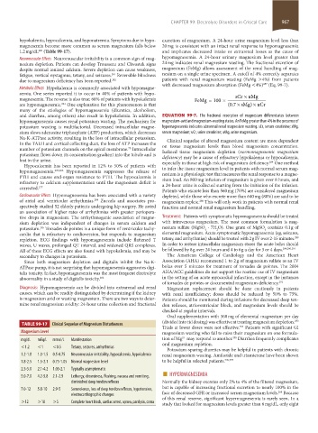
taBLE 99-17 Clinical Sequelae of Magnesium Disturbances (divided into tid dosing) was effective at treating magnesium depletion.
Trials at lower doses were not effective. Patients with significant GI
294
Magnesium Level magnesium wasting who fail to raise their magnesium on one formula-
295
2+
mg/dL mEq/L mmol/L Manifestation tion of Mg may respond to another. Diarrhea frequently complicates
oral magnesium repletion.
<1.2 <1 <0.5 Tetany, seizures, arrhythmias Potassium-sparing diuretics may be helpful in patients with chronic
1.2-1.8 1.0-1.5 0.5-0.75 Neuromuscular irritability, hypocalcemia, hypokalemia renal magnesium wasting. Amiloride and triamterene have been shown
1.8-2.5 1.5-2.1 0.75-1.05 Normal magnesium level to be helpful in selected patients. 296,297
2.5-5.0 2.1-4.2 1.05-2.1 Typically asymptomatic ■
5.0-7.0 4.2-5.8 2.1-2.9 Lethargy, drowsiness, flushing, nausea and vomiting, HYPERMAGNESEMIA
diminished deep tendon reflexes Normally the kidney excretes only 2% to 4% of the filtered magnesium,
7.0-12 5.8-10 2.9-5 Somnolence, loss of deep tendon reflexes, hypotension, but is capable of increasing fractional excretion to nearly 100% in the
268
electrocardiographic changes face of decreased GFR or increased serum magnesium levels. Because
>12 >10 >5 Complete heart block, cardiac arrest, apnea, paralysis, coma of this renal reserve, significant hypermagnesemia is rarely seen. In a
study that looked for magnesium levels greater than 6 mg/dL, only eight
section08.indd 967 1/19/2015 11:09:54 PM