
Page 1393 - Hall et al (2015) Principles of Critical Care-McGraw-Hill
P. 1393
966 PART 8: Renal and Metabolic Disorders
nearly zero in the presence of hypermagnesemia or reduced GFR (ie, all TABLE 99-16 Etiologies of Hyper- and Hypomagnesemia (Continued)
of the filtered magnesium is excreted). In response to magnesium deple-
tion or decreased intake, the fractional resorption of Mg can rise to Hypomagnesemia
2+
99.5% in order to minimize urinary losses. Increased tubular flow
■ HYPOMAGNESEMIA Osmotic diuresis
Hypomagnesemia is common, occurring in approximately 12% of hos- Diabetes types I and II
pitalized patients. Among ICU patients, the prevalence of hypomagne- Hyperaldosteronism e
257
semia ranges from 11% to 65%. 258-260 Hypomagnesemia frequently goes Volume expansion
undetected. In a prospective study, 47% of patients undergoing clinical Diabetic ketoacidosis
blood testing for electrolyte concentrations had hypomagnesemia, but
physicians ordered magnesium levels in only 10% of these patients. 261 Tubular dysfunction
Recovery from acute
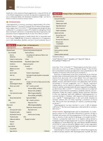
Etiologies: Hypomagnesemia is nearly always due to increased renal
or GI losses (Table 99-16). GI losses or malabsorption of magnesium tubular necrosis
occur with steatorrhea, diarrhea, and short bowel syndrome (loss of Recovery from obstruction
Recovery from
transplantation
TABLE 99-16 Etiologies of Hyper- and Hypomagnesemia
Congenital renal magnesium
Hypomagnesemia Hypermagnesemia wasting
Extrarenal causes Decreased renal excretion of magnesium Bartter syndrome (one-third
Gastrointestinal Renal insufficiency of cases)
Diarrhea Any etiology with a glomerular filtration rate Gitelman syndrome (universal)
Steatorrhea <10 mL/min Data from these references:
264 c
340 b
341 d
267 f
342 e
Congenital malabsorption Lithium a Galland ; Hessov et al ; Lipner ; Papazachariou et al ; Massry et al ; Sutton and
268 g
Domrongkitchaiporn ; Eshleman et al 343
Protein calorie malnutrition Hypocalciuria, hypercalcemia f
Alcoholism Magnesium ingestion
Enteral nutrition Parenteral more than 75 cm of bowel). 262-264 Hypomagnesemia has been associ-
Inflammatory bowel Dosing error ated with concurrent use of PPI and diuretic therapy. The US FDA
265
disease a has recommended monitoring magnesium levels periodically for the
Gastric suction Treatment of preeclampsia duration of treatment with proton-pump inhibitors. 266
Renal loss of magnesium occurs most prominently in any situation
Vomiting Treatment of torsades de pointes or myocardial in which there is increased tubular flow. Intravenous fluids or osmotic
infarction
diuresis from glucosuria will increase tubular flow and magnesium
Short bowel syndrome b Oral wasting. Loop, thiazide, and osmotic diuretics, recovery from acute
267
Sprue Damage to the intestinal lining may increase Mg tubular necrosis, and relief of urinary tract obstruction have all been
Intestinal bypass for obesity c absorption documented to increase magnesium loss. 268-270 Specific magnesium
wasting defects can be induced by tubular toxins. Cisplatin, ampho-
2+
Chronic pancreatitis d Mg -containing antacids
tericin B, and the aminoglycosides all cause magnesium wasting inde-
Skin Gaviscon [Al(OH) and MgCO ] pendent of any effect on GFR. 271-273 Gitelman syndrome is a congenital
3 3
Burns Mylanta (CaCO and MgCO ) syndrome characterized by hypokalemia, metabolic alkalosis, and nor-
3 3 motension. Unlike the similar condition Bartter syndrome, Gitelman
Toxic epidermal necrolysis Milk of magnesia [Mg(OH) ]
2 is often not diagnosed until early adulthood. Hypomagnesemia is a
Bone Maalox [Al(OH) and Mg(OH) ] universal finding in Gitelman, with magnesium levels typically just
3 2
274
Hungry bone syndrome Epsom salts (MgSO ) over 1 mg/dL. Hypomagnesemia is also particularly common in
4
2+
Other Mg -containing cathartics alcoholic patients, with one study reporting a prevalence of almost
30%. This results from the interplay of a number of pathophysiologi-
Pancreatitis Magnesium citrate cal factors. 275
Renal causes Milk of magnesia [Mg(OH) ] Hypomagnesemia has been reported to occur in 40% of patients
2
276
Drugs Magnesium-containing enemas with burns. The decreased magnesium is due primarily to exudative
skin losses. 277
Aminoglycoside toxicity Magnesium citrate
Pentamidine toxicity Aspiration Clinical Sequelae: Hypomagnesemia may be asymptomatic. In a retro-
spective review of 1576 consecutive admissions to a geriatric facility in
Amphotericin B toxicity Dead Sea near drowning Scotland, 169 patients with hypomagnesemia (≤1.6) showed no differ-
278
Thiazide diuretics Other ence in duration of stay, survival to discharge, or 6-month survival.
Calcineurin inhibitors Theophylline toxicity g However, a prospective study done in an inpatient setting showed a
tremendous impact of hypomagnesemia on survival. Though there was
Foscarnet
no difference in Acute Physiology, Age, and Chronic Health Evaluation
Cisplatin (APACHE) II scores at admission, patients with a serum magnesium
Loop of Henle level <1.5 mg/dL had a dramatically higher mortality rate than patients
with normal magnesium (31% vs. 22%). 279
Loop diuretics
Determining the clinical consequences of isolated hypomagnesemia
Hypercalcemia (Continued) is difficult because patients with hypomagnesemia typically also have
section08.indd 966 1/14/2015 8:28:23 AM