
Page 1392 - Hall et al (2015) Principles of Critical Care-McGraw-Hill
P. 1392
CHAPTER 99: Electrolyte Disorders in Critical Care 965
246
Etiologies cardiac arrhythmias. The risk of calcification increases as the calcium-
2
2
Increased Intake of Phosphorus The ability to maintain phosphorus balance phosphorus product (calcium times phosphorus) rises above 70 mg /dL .
in the face of massive phosphorus loads (4000 mg/d) depends on the In patients with end-stage renal disease, empiric data have shown
phosphorus load being spread over time. Sudden loads can overwhelm decreased mortality in patients with a calcium-phosphorus product less
renal phosphate clearance, resulting in hyperphosphatemia. Phosphorus than 52. In the same study, isolated hyperphosphatemia also predicted
loads can be exogenous or endogenous (Table 99-15). Exogenous intake increased mortality. 247
can be from diet, phosphate enemas, or parenteral sources. Fleet enemas Treatment: Hyperphosphatemia in patients with intact renal func-
contain 130 mg (4.15 mmol) of phosphorus per milliliter. Dietary tion is usually transient and self-correcting. Infusing saline to induce
intake of phosphorus can be enhanced by vitamin D toxicity. Calcitriol natriuresis can enhance renal clearance of phosphorus. Acetazolamide
enhances gut absorption of phosphorus and the associated hypercal- can increase renal clearance by blocking phosphate resorption in the
cemia, along with the increased calcitriol, suppresses PTH, decreasing proximal tubule. 248
renal phosphorus clearance. If there is decreased renal function or symptomatic hypocalcemia,
Endogenous sources of phosphate are due to release of intracellular dialysis is essential. Twenty to thirty millimoles of phosphorus are
phosphorus from cell death or transcellular distribution. Patients who removed with a 4-hour dialysis session. Continuous renal replacement
present with diabetic ketoacidosis are usually hyperphosphatemic strategies have been shown to provide better control of hyperphospha-
despite decreased total body phosphorus. This is due to the lack of temia and hypocalcemia. 249
244
insulin decreasing the movement of phosphorus into cells and metabolic In tumor lysis syndrome, the use of sodium bicarbonate to alkalinize
acidosis slowing phosphorus consuming glycolysis. Tumor lysis syn- the urine can be detrimental. Alkalinization has been used to increase
drome is due to destruction of large bulky tumors with chemotherapy solubility of uric acid in the urine; however, urinary phosphorus solu-
or radiation therapy. The tumor cells release phosphorus, potassium, bility decreases with higher urine pH. The use of sodium bicarbonate
and purines (metabolized to uric acid). Acute renal failure from urate predisposes to renal calcium deposition. In addition, raising the pH
nephropathy can exacerbate the electrolyte abnormalities. For more exacerbates the ionized hypocalcemia found in tumor lysis syndrome.
information, see the discussion in “Hyperkalemia”, above. The use of allopurinol and uricase prevents hyperuricemia, eliminating
Decreased Renal Clearance of Phosphorus Since the kidney is the primary means the need for alkalinization.
of excreting phosphorus, renal failure of any etiology is associated Phosphate binders are regularly used in patients with chronic renal
with hyperphosphatemia. The kidney maintains phosphorus balance failure to reduce absorption of dietary phosphorus. Though they primar-
by filtering serum phosphorus and then adjusting the fractional resorp- ily act to decrease absorption of dietary phosphorus, they have a small
tion of phosphorus via PTH. In some cases, the kidneys fail to excrete but measurable ability to reduce phosphorus in patients not ingesting
phosphorus despite adequate GFR. The primary cause of this is hypopara- additional phosphorus. Patients with acute hyperphosphatemia should
250
thyroidism due to removal of the parathyroids or other neck surgery. In have a low-phosphorus diet and be started on phosphorus binders
the former, the hypoparathyroidism is permanent, while in the latter it is (ie, magnesium or calcium salts, lanthanum carbonate, or sevelamer).
usually a temporary stunning of the gland. Other causes of hypoparathy-
roidism are discussed under etiologies of hypocalcemia (see Table 99-8).
Clinical Sequelae: The primary clinical consequence of hyperphospha- MAGNESIUM
temia is hypocalcemia and its metabolic manifestations. Increased ■ METABOLISM
serum phosphorus binds ionized calcium, lowering the biologically
active fraction of calcium. 245 Magnesium is the second most prevalent intracellular cation. It is a
Severe hyperphosphatemia can result in metastatic calcification in critical cofactor in any reaction powered by ATP, so deficiency of this
soft tissues. In rare cases, this may contribute to acute renal failure or ion can have dramatic effects on metabolism. Magnesium also acts as
a calcium channel antagonist and plays a key role in the modulation
of any activity governed by intracellular calcium (eg, muscle contrac-
tion and insulin release). The atomic weight of magnesium is 24.3.
251
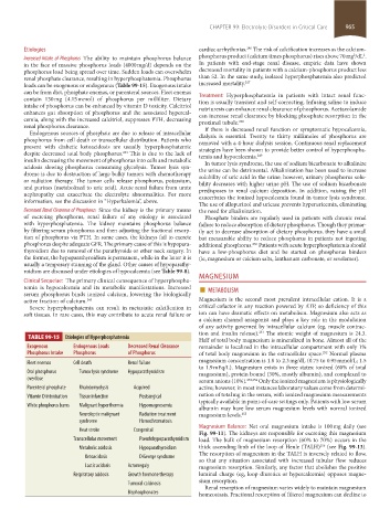
TABLE 99-15 Etiologies of Hyperphosphatemia
Half of total body magnesium is mineralized in bone. Almost all of the
Exogenous Endogenous Loads Decreased Renal Clearance remainder is localized in the intracellular compartment with only 1%
Phosphorus Intake Phosphorus of Phosphorus of total body magnesium in the extracellular space. Normal plasma
252
magnesium concentration is 1.8 to 2.3 mg/dL (0.75 to 0.95 mmol/L; 1.5
Fleet enemas Cell death Renal failure
to 1.9 mEq/L). Magnesium exists in three states: ionized (60% of total
Oral phosphorus Tumor lysis syndrome Hypoparathyroidism magnesium), protein bound (30%, mostly albumin), and complexed to
overdose serum anions (10%). 253,254 Only the ionized magnesium is physiologically
Parenteral phosphate Rhabdomyolysis Acquired active; however, in most instances laboratory values come from determi-
Vitamin D intoxication Tissue infarction Postsurgical nation of total mg in the serum, with ionized magnesium measurements
typically available in point-of-care settings only. Patients with low serum
White phosphorus burns Malignant hyperthermia Hypomagnesemia
albumin may have low serum magnesium levels with normal ionized
Neuroleptic malignant Radiation treatment magnesium levels. 255
syndrome Hemochromatosis
Magnesium Balance: Net oral magnesium intake is 100 mg daily (see
Heat stroke Congenital
Fig. 99-11). The kidneys are responsible for excreting this magnesium
Transcellular movement Pseudohypoparathyroidism load. The bulk of magnesium resorption (60% to 70%) occurs in the
256
Metabolic acidosis Hypoparathyroidism thick ascending limb of the loop of Henle (TALH) (see Fig. 99-13).
The resorption of magnesium in the TALH is inversely related to flow,
Ketoacidosis DiGeorge syndrome
so that any situation associated with increased tubular flow reduces
Lactic acidosis Acromegaly magnesium resorption. Similarly, any factor that abolishes the positive
Respiratory acidosis Growth hormone therapy luminal charge (eg, loop diuretics or hypercalcemia) opposes magne-
sium resorption.
Tumoral calcinosis
Renal resorption of magnesium varies widely to maintain magnesium
Bisphosphonates homeostasis. Fractional resorption of filtered magnesium can decline to
section08.indd 965 1/14/2015 8:28:23 AM