
Page 1789 - Hall et al (2015) Principles of Critical Care-McGraw-Hill
P. 1789
1258 PART 11: Special Problems in Critical Care
hemorrhage. Uterine atony is associated with uterine overdistension, pla-
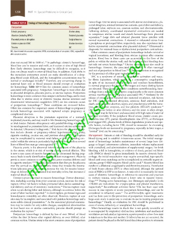
TABLE 127-5 Etiology of Hemorrhagic Shock in Pregnancy
cental abruption, retained intrauterine contents, quick labor and delivery,
Antepartum Postpartum prolonged labor, oxytocin use, cesarean section, and chorioamnionitis.
Ectopic pregnancy Uterine atony Following delivery, coordinated myometrial contractions are needed
to compresses uterine vessels and stanch hemorrhage from placental
Abortion (including RPOC) Retained placenta
separation. Large clots and retained placental tissue interfere with
28
Placental previa or abruption Surgical trauma normal myometrial contractions. A stunned or exhausted uterus from
Uterine rupture in VBAC Uterine inversion a precipitous or prolonged labor, respectively, may also experience inef-
38
Trauma DIC fective myometrial contractions after placental delivery. Ultrasound is
diagnostic for retained tissue or dysfunctional postpartum contractions.
DIC, disseminated intravascular coagulation; RPOC, retained products of conception; VBAC, vaginal birth Other common causes of postpartum hemorrhage include cervical or
https://kat.cr/user/tahir99/
after caesarian section.
vaginal lacerations, and bleeding from uterine incisions after cesarean
section. Blood loss in these cases can accumulate in the floor of the
does not exceed 500 to 1000 mL. In pathologic obstetric hemorrhage, pelvis or within the uterine wall, and the lack of evident bleeding does
30
31
blood loss can be massive and swift, as it occurs at sites of high blood not rule out severe hemorrhage. Uterine inversion may also result in
flow. Early obstetric hemorrhage may be difficult to recognize, as it hemorrhage. However, the associated hypotension is often vasovagal
38,39
does not always result in external blood loss. In addition, fluid shifts in and out of proportion to blood loss. Uterine inversion is recognized
the immediate postpartum period can make identification of a drop- by the presence of a blue-gray vaginal protrusion.
ping blood count difficult, and the hemoglobin concentration may be DIC is a syndrome of systemic coagulation activation and vascu-
normal or unchanged initially. Therefore, any concerning change in lar fibrin deposition, which results in a consumptive coagulopathy.
28
maternal heart rate or blood pressure should prompt an evaluation In spite of an increased plasma volume and resultant hemodilution,
for hemorrhage. Table 127-5 lists the common causes of hemorrhage in normal pregnancy the levels of fibrinogen and many clotting factors
associated with pregnancy. Antepartum hemorrhage is most often due are elevated. These hypercoagulable conditions notwithstanding, hem-
to placental abruption, placenta previa, or uterine rupture. Postpartum orrhage from a massive consumptive coagulopathy is the most common
40
hemorrhage is more common than antepartum hemorrhage, and is most serious manifestation of pregnancy-associated DIC. Mediated by the
often due to uterine atony or obstetric trauma; uterine inversion and release of procoagulant material into maternal circulation, risk factors
disseminated intravascular coagulation (DIC) are less common causes for DIC include placental abruption, amniotic fluid embolism, fetal
of postpartum hemorrhage. These conditions are reviewed below. death, saline solution abortion, sepsis, and preeclampsia with the hemo-
1,3
40
Other less common but important causes of hemorrhage in pregnancy lysis, elevated liver enzymes, and low platelets (HELLP) syndrome.
include hemorrhage associated with ruptured ectopic pregnancy or DIC may occur before or after delivery, and the onset is often abrupt.
complicated abortion. The course may be fulminant and associated with high rates of maternal
Placental abruption is the premature separation of a normally and fetal mortality. If the peripheral blood smear, platelet count, pro-
implanted placenta, and may result in life-threatening hemorrhage and/ thrombin time (PT), partial thromboplastin time (PTT), or fibrinogen
or fetal demise. Patients often present with painful bleeding, which may level suggest DIC, plasma levels of fibrin degradation products and spe-
be misdiagnosed as premature labor, and increased uterine activity may cific factors, including factor VIII, should be measured. As circulating
be detected. Ultrasound is diagnostic. Risk factors for placental abrup- fibrinogen levels are increased in pregnancy, especially in later stages, a
31
40
tion include chronic or pregnancy-related hypertension, high parity, “normal” level can be concerning.
cigarette smoking, cocaine use, and previous abruption. 32-35 Abruption Management: Patients at risk of bleeding should be identified early for
may be complicated by maternal renal failure or DIC. Bleeding con- blood typing and to establish intravenous access. The initial manage-
3,34
cealed within the uterus is particularly high risk for fetal death as several ment of hemorrhage includes maintenance of several large-bore (16-
liters of blood loss may go unrecognized. 31,34 gauge or larger) intravenous catheters, immediate volume replacement
Placenta previa is the abnormal inferior attachment of the placenta with crystalloid, and administration of supplemental oxygen. For brisk
in the uterus, which is at risk of tearing during cervical dilation. This bleeding, a fall in hemoglobin, or evidence of shock, packed red blood
is now a rare cause of massive hemorrhage as ultrasound during preg- cells (PRBCs) should be given immediately. In massive obstetric hem-
nancy leads to early identification and expectant management. Placenta orrhage, the initial resuscitation may require unmatched, type-specific
previa is more common in multiparas with prior cesarean delivery and blood until cross-matching can be accomplished; in critically urgent sit-
in cigarette smokers. 1,32,36 Vaginal examination that disrupts the placenta uations, group O RhD-negative blood can be used. Massive blood loss
41
over the cervical os, and trophoblastic tissue that invades the myome- results in a dilutional coagulopathy and thrombocytopenia. Transfusion
trium (placenta previa et accreta) increase the risk for massive hemor- of fresh frozen plasma (FFP) is often indicated, although the optimal
rhage at delivery. The associated fetal mortality is low, but increases if ratio of PRBCs to FFP is not known. A ratio of 6:1 is reasonable for most
33
maternal shock occurs. cases of obstetric hemorrhage; in reference to outcomes and practices
Uterine rupture can result in massive hemorrhage. Uterine abnormal- in military trauma, some advocate a lower ratio in massive hemor-
ities, including scarring from prior cesarean section, increase the risk of rhage. 42,43 During active hemorrhage, low fibrinogen and platelet levels
rupture. Other risk factors include protracted labor, device-assisted vag- <50,000 are indications for cryoprecipitate and platelet transfusions,
inal delivery, and use of uterotonic medications. Uterine rupture most respectively. Recombinant activated factor VIIa has been used with
42
36
often occurs during labor and delivery, although occurrence before the success in case reports of severe postpartum hemorrhage, and can be
onset of labor has been reported. In overt rupture, peritoneal signs and considered in refractory cases. The antifibrinolytic agent tranexamic
37
42
hemodynamic instability are often observed. However, rupture at scar acid has been used in the prevention of postpartum hemorrhage, and a
sites may be incomplete, and associated with painless hemorrhage and a large-scale study is underway to evaluate its use in treating postpartum
more subtle clinical presentation. As the associated physical examina- hemorrhage. Finally, an evaluation for DIC should be performed in
42
31
tion may be notable for only subtle changes, unexplained abnormalities cases of severe, refractory, or unexplained hemorrhage.
in fetal heart rate or uterine contractility patterns should prompt an When hemorrhage results in shock that is not quickly reversible or
evaluation for rupture. 31 is accompanied by respiratory dysfunction, intubation and mechanical
Postpartum hemorrhage is defined by loss of over 500 mL of blood ventilation are indicated as hypoxemia superimposed on a low-flow state
within the first 24 hours after vaginal delivery, or over 1000 mL after is injurious to the fetus and mother. If delivery has not yet occurred, the
cesarean section. Uterine atony is the most common cause of postpartum patient should be placed in the left lateral decubitus position to attenuate
section11.indd 1258 1/19/2015 10:52:21 AM