
Page 1824 - Hall et al (2015) Principles of Critical Care-McGraw-Hill
P. 1824
CHAPTER 129: Dermatologic Conditions 1293
erythematous plaques, which may coalesce to form “lakes of pus.” often accompanied by fever, chills, and lymphadenopathy, follow.
Nails, palms, and soles are often affected. Fever, hypocalcemia, and The percutaneous absorption barrier is lost and blood flow to the
leukocytosis may accompany the outbreak. Precipitating factors include skin increases, which may lead to serious complications such as
drugs, infection, pregnancy, exertion, and menstruation. The differential hypoalbuminemia, peripheral edema, loss of muscle mass, and
diagnosis in a patient with diffuse pustules and fever includes infection, high-output cardiac failure, with as much as 8% of the total cardiac
drug reaction, acute generalized exanthematous pustulosis, pustular output being directed at the inflamed cutaneous vasculature. Causes
psoriasis, and subcorneal pustular dermatosis. 77,78 of exfoliative dermatitis include inflammatory conditions, drug
Psoriasis may be treated initially with a variety of topical medications, eruptions, cutaneous T-cell lymphoma, and systemic neoplasms
either as monotherapy or in combination. These include corticosteroids, (Table 129-15). Common inflammatory conditions include psoria-
calcipotriene, tar, anthralin, and topical retinoids. Systemic drugs are sis, atopic dermatitis, contact dermatitis, and pityriasis rubra pila-
reserved for extensive or disabling disease and include methotrexate, ris. Antiepileptic medications (carbamazepine, phenobarbital, or
cyclosporine, and oral retinoids. Ultraviolet (UV) light therapy, which phenytoin), antihypertensive medications (captopril or chlorothia-
has been a mainstay of treatment for years, is often used in combination zide), antibiotics (cephalosporins, dapsone, isoniazid, or mino-
with topical or systemic agents. This includes natural sunshine, broad- cycline), and calcium channel blockers have been associated with
band UVB (280-20 nm), UVA (320-400 nm), and a single-wavelength exfoliative erythroderma. Lymphomas and hematologic malignancies
80
light narrowband UVB (311 nm). Psoralen, a photosensitizer, is used are common systemic neoplasms that may cause erythroderma. Sézary
to potentiate the UVA effects in a regimen called PUVA. Acitretin, a syndrome is the leukemic form of cutaneous T-cell lymphoma, which is
systemic retinoid, may be used alone or in combination with photo- characterized by circulating atypical lymphocytes with hyperconvoluted
therapy. Biologic therapy that targets specific cytokines and intercellular nuclei (Sézary cells). These atypical cells are identified on blood smear or
adhesion molecules has recently been introduced. Biologics include in skin biopsies. Internal malignancies occasionally cause erythroderma.
TNF-α inhibitors (etanercept, adalimumab, infliximab) and T-cell The erythrodermic patient should be monitored with specific
modulators (alefacept, efalizumab). Acitretin, narrowband UVB, PUVA, attention to fluid and electrolyte balance, temperature regulation, and
methotrexate, and cyclosporine have been used effectively in the treat- nutritional status. Skin biopsy may help identify the underlying cause of
ment of generalized pustular psoriasis. 79 the dermatosis and thus direct specific treatment. Initial management
■ ERYTHRODERMA includes the use of medium potency topical steroid ointments covered
with a bland ointment, such as zinc oxide ointment and wrapped with
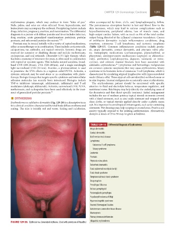
Erythroderma or exfoliative dermatitis (Fig. 129-20) is a descriptive term clean cloths, or topical steroids applied directly under a plastic sauna
for a clinical condition characterized by total body diffuse erythema and suit. It is important to avoid topical irritant agents, such as tar-containing
scaling. The skin is initially red and warm. Scaling and exfoliation, ointments. Wet dressings may help weeping or crusted areas. Pruritus and
anxiety typically respond to the sedating antihistamines. Alternatively,
doxepin at doses of 25 to 50 may be given at bedtime.
TABLE 129-15 Differential Diagnosis of Erythroderma
Atopic dermatitis
Contact dermatitis
Graft-versus-host disease
Lymphoma:
Cutaneous T-cell lymphoma
Sézary syndrome
Leukemia
Psoriasis
Pityriasis rubra pilaris
Seborrheic dermatitis
Toxic epidermal necrolysis (early)
Toxic shock syndrome
Streptococcal toxic shock syndrome
Drug eruption
Pemphigus foliaceus
Bullous pemphigoid
Paraneoplastic pemphigus
Papuloerythroderma of Ofuji
Hypereosinophilic syndrome
Crusted (Norwegian) Scabies
Autoimmune connective tissue disease
Mastocytosis
Primary immunodeficiencies
FIGURE 129-20. Erythroderma. Generalized erythema. (Used with permission of VisualDx.) Idiopathic erythroderma
section11.indd 1293 1/19/2015 10:54:16 AM